
Joint Dislocation and Subluxation
Key points
- Definitions: dislocation is complete loss of congruity between two articular surfaces; subluxation is partial loss, with some contact remaining.
- Before any reduction: document neurovascular status, give adequate analgesia or procedural sedation, and obtain a pre-reduction radiograph unless circulation is critically compromised.
- Anterior shoulder dislocation: by far the commonest, from forced abduction, external rotation and extension. Check axillary nerve function - sensation over the regimental badge area and deltoid contraction - before and after reduction.
- Posterior shoulder dislocation: classically follows a seizure or electric shock. Easily missed on a single AP film; the arm is held adducted and internally rotated and cannot be externally rotated.
- Hip dislocation: usually posterior, from a dashboard injury or after hip replacement. The leg is shortened, flexed, adducted and internally rotated - the opposite position to a neck of femur fracture.
- Hip dislocation is time-critical: reduce within 6 hours to minimise the risk of avascular necrosis of the femoral head, and assess for sciatic nerve injury.
- Pulled elbow: radial head subluxation in young children after a pulling force on the arm. Usually reduced clinically without imaging when the history is classic.
- Patellar dislocation: typically lateral, in young hypermobile females, after a twisting movement on a planted foot. Assess with the apprehension test and consider an osteochondral fracture.
Introduction
A dislocation is complete loss of congruity between the two articular surfaces of a joint, such that they are no longer in contact at all. A subluxation is a partial dislocation, where some contact between the articular surfaces remains.1 Both occur when a force applied to a joint exceeds the restraining capacity of its capsule, ligaments and surrounding muscles.
Dislocations matter for reasons beyond the obvious deformity and pain. The joint capsule, ligaments and labrum are torn, predisposing to recurrent instability. Nearby nerves and vessels, which often run close to the joint, can be stretched, compressed or torn by the displaced bone. A fracture frequently accompanies the dislocation, and the combination - a fracture-dislocation - is more unstable and harder to treat than either injury alone.
This is a heavily examined area because the diagnosis is often made from the mechanism and the position of the limb alone, and because a handful of associated injuries - axillary nerve palsy in anterior shoulder dislocation, sciatic nerve injury and avascular necrosis in hip dislocation - are exactly the kind of detail that separates a pass from a distinction.
General principles of management
The same sequence applies to almost every dislocation, regardless of the joint involved, and is a reliable structure for an exam answer or an acute presentation.
- Analgesia first. Reduction is painful, and attempting it without adequate analgesia or procedural sedation causes unnecessary suffering and makes the muscle spasm that resists reduction worse. Options range from intravenous opioids and Entonox to formal procedural sedation, depending on the joint and the setting.8
- Assess and document neurovascular status before any reduction attempt. Record distal pulses, capillary refill, and the sensory and motor function of every nerve at risk for that joint. This is the single most important thing to write in the notes, because it establishes whether a deficit found afterwards was caused by the dislocation or by the reduction.
- Obtain a pre-reduction radiograph to confirm the direction of dislocation and identify any associated fracture, since this changes the reduction technique and the urgency. The exception is a grossly deformed dislocation that is compromising the overlying skin or the circulation, where clinical urgency overrides imaging and reduction should not be delayed.
- Reduce using an appropriate technique, with muscle relaxation from analgesia or sedation making this considerably easier.
- Reassess and document neurovascular status immediately after reduction. A new deficit after reduction that was not present before points to iatrogenic nerve stretch or entrapment and needs urgent discussion with orthopaedics.
- Obtain a post-reduction radiograph to confirm the joint is congruent and to look for a fracture that may have been caused, or unmasked, by the reduction.
- Immobilise the joint appropriately - typically a sling for the shoulder, a splint or brace for the elbow or patella - for a period balanced against the risk of stiffness.
- Arrange follow-up and physiotherapy, since rehabilitation of the surrounding muscles reduces the risk of recurrent instability, and referral to orthopaedics for recurrent or complex dislocations.
Shoulder (glenohumeral) dislocation
The glenohumeral joint is inherently unstable - a large humeral head articulating with a shallow glenoid, trading stability for the greatest range of motion of any joint in the body - which is why it is the most commonly dislocated large joint, and by far the most heavily examined.
Anterior dislocation
Anterior dislocation accounts for around 95% of shoulder dislocations.5 The typical mechanism is a force that combines abduction, external rotation and extension of the arm - classically a fall onto an outstretched, abducted arm, or a direct blow to the back of the shoulder that drives the humeral head forward.
Clinically, the normal rounded contour of the shoulder is lost, replaced by a squared-off appearance as the deltoid falls in over the empty glenoid. The arm is held in slight abduction and external rotation, supported by the other hand, and any attempt at movement is guarded and painful. A fullness may be palpable anteriorly where the humeral head now lies.
Two structural lesions are frequently associated and worth naming specifically:
- Bankart lesion - a tear of the anteroinferior glenoid labrum, sometimes with an avulsed fragment of glenoid bone (a bony Bankart). This removes part of the static restraint that normally deepens the socket.
- Hill-Sachs lesion - a compression (impaction) fracture of the posterolateral humeral head, caused as it is forced against the anterior rim of the glenoid at the moment of dislocation.
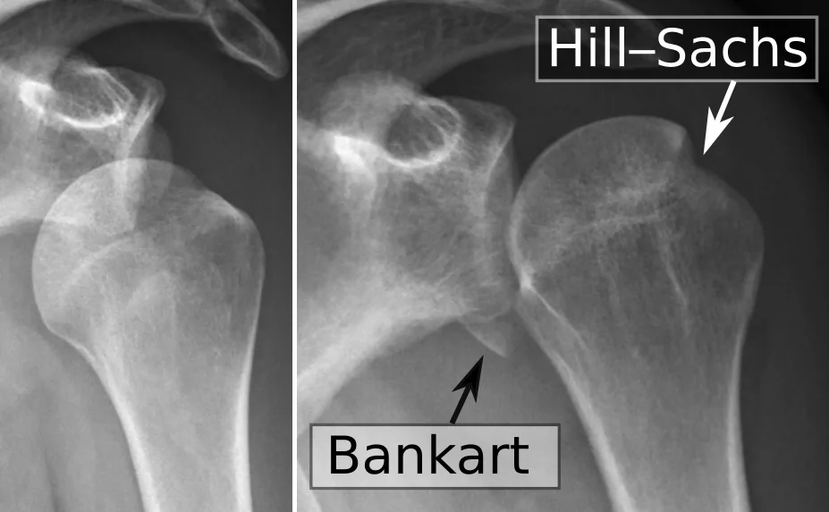
Both lesions are relevant chiefly because of recurrent instability. Younger patients, particularly men under 25 having their first traumatic dislocation, have a substantially higher risk of a further dislocation, since the torn labrum and capsule heal poorly without surgical repair. Older patients are more likely to sustain a rotator cuff tear instead, and are less likely to redislocate.3
Posterior dislocation
Posterior dislocation is much rarer, making up only a small fraction of shoulder dislocations, but is disproportionately tested because it is disproportionately missed.
The arm is held adducted and internally rotated, and the patient is unable to externally rotate it - a useful bedside discriminator, since anterior dislocation holds the arm externally rotated. A standard AP shoulder radiograph can look deceptively normal because the humeral head remains roughly overlapping the glenoid on this view (the 'light bulb sign' of a fixed internally rotated head is a subtle clue). An axillary view or scapular Y view, or a CT scan where doubt remains, is needed to confirm the diagnosis.
Reduction and aftercare
Several closed reduction techniques exist for anterior dislocation, chosen according to local practice, patient build and available analgesia/sedation:
- Traction-countertraction - steady longitudinal traction on the arm while an assistant applies countertraction across the chest with a sheet, gradually overcoming muscle spasm
- Kocher's method - a sequence of external rotation, adduction and internal rotation with the elbow flexed to 90 degrees; effective but carries a higher risk of fracture if performed forcefully
- Hippocratic method - traction with the clinician's foot placed in the axilla, now used less often given the risk of neurovascular injury from the counter-pressure
- Scapular manipulation - performed with the patient prone, rotating the inferior scapular tip medially to realign the glenoid with the humeral head
After successful reduction, the arm is placed in a sling for comfort, followed by physiotherapy to restore range of movement and strengthen the rotator cuff and scapular stabilisers. Patients with recurrent dislocation, or a first dislocation in a young, active patient at high risk of recurrence, are referred for consideration of surgical stabilisation.
Hip dislocation
The hip is an inherently stable, deep ball-and-socket joint, so dislocation requires a high-energy force and should always prompt a search for associated injuries elsewhere, particularly in polytrauma.
Posterior dislocation
Roughly 90% of traumatic hip dislocations are posterior.6 The classic mechanism is a dashboard injury: a road traffic collision in which the knee strikes the dashboard while the hip is flexed, driving the femoral head backwards out of the acetabulum. It is also seen after total hip replacement, where the prosthetic femoral head can dislocate posteriorly with far less force, particularly with certain hip flexion and internal rotation movements.
Anterior dislocation
Anterior dislocation is much less common and results from a force that combines abduction and external rotation of the hip, for example a fall from a height landing with the legs abducted. The leg lies abducted and externally rotated, the opposite posture to a posterior dislocation, and the femoral head may be palpable anteriorly in the groin. The femoral neurovascular bundle is at risk given its proximity.
Elbow dislocation
The elbow is the second most commonly dislocated large joint in adults, and the most common in children. The great majority are posterior dislocations, typically from a fall onto an outstretched hand with the elbow extended, which drives the ulna and radius backwards relative to the humerus.
Clinically, the elbow is held fixed in flexion, with an obvious deformity and swelling, and the normal triangular relationship of the olecranon and the two epicondyles is disrupted (useful for distinguishing dislocation from a supracondylar fracture, where this relationship is preserved).
The neurovascular bundle crossing the elbow is at real risk of injury and must be assessed carefully before and after reduction:
- Brachial artery - check the radial pulse and capillary refill; the artery can be stretched, kinked or, rarely, transected
- Median nerve - sensation over the palmar radial three and a half digits, and thumb opposition/flexion; particularly at risk with an associated medial epicondyle fracture in children, where the nerve can become entrapped in the joint
- Ulnar nerve - sensation over the little finger and medial one and a half digits, and finger abduction/adduction
- Radial nerve - sensation over the dorsal first web space, and wrist/finger extension
Reduction is achieved with steady longitudinal traction and countertraction, correcting any coronal (varus/valgus) displacement first, under adequate analgesia. Following reduction, the elbow is immobilised in a splint, with early mobilisation guided by physiotherapy to reduce the substantial risk of stiffness that follows elbow injuries of any kind.
Patellar dislocation
Patellar dislocation is typically lateral, and is characteristically seen in young, hypermobile, often female patients, though it also occurs after direct trauma in anyone. The usual mechanism is a twisting movement with the foot planted and the knee slightly flexed, generating a strong contraction of quadriceps that pulls the patella laterally out of the trochlear groove, sometimes with a preceding valgus knee position.
The patient reports the knee 'giving way' or feeling like it popped out, with immediate pain and swelling, and a visibly displaced patella if it has not already spontaneously reduced (which happens surprisingly often, particularly on straightening the knee, before the patient is even seen). A joint effusion (haemarthrosis) is common.
The apprehension test is the key examination finding: with the knee slightly flexed, the examiner pushes the patella laterally, and a patient who has previously dislocated will actively resist and show visible apprehension or contract their quadriceps to guard against it. This test is deliberately provocative and should be performed gently.
An important complication to actively look for is an osteochondral fracture, produced as the patella shears against the lateral femoral condyle during dislocation or relocation; a fragment of articular cartilage and bone can shear off and become a loose body within the joint, and may need surgical removal or fixation. Recurrent instability is common, particularly where there is underlying trochlear dysplasia, patella alta, or generalised ligamentous laxity, and recurrent dislocators are referred for consideration of physiotherapy-led quadriceps rebalancing or, in refractory cases, surgical stabilisation (such as medial patellofemoral ligament reconstruction).4
Summary of key joints
| Joint | Typical direction | Classic mechanism | Clinical findings | Nerve/vessel at risk |
|---|---|---|---|---|
| Shoulder | Anterior (~95%) | Fall on outstretched, abducted arm; direct blow to posterior shoulder | Loss of shoulder contour, arm in slight abduction and external rotation | Axillary nerve |
| Shoulder | Posterior | Seizure, electric shock | Arm adducted and internally rotated, unable to externally rotate | Axillary nerve |
| Hip | Posterior (~90%) | Dashboard injury (RTC); post-hip replacement | Leg shortened, flexed, adducted, internally rotated | Sciatic nerve; femoral head blood supply (AVN risk) |
| Hip | Anterior | Fall with hip abducted and externally rotated | Leg abducted and externally rotated | Femoral neurovascular bundle |
| Elbow | Posterior | Fall on outstretched hand | Fixed flexion, deformity, disrupted triangular bony relationship | Brachial artery; median, ulnar and radial nerves |
| Patella | Lateral | Twisting movement on a planted foot | Visible/palpable displacement (may spontaneously reduce), positive apprehension test | None characteristic; risk of osteochondral fracture |
Complications
Complications are broadly shared across dislocated joints, though the relative risk of each varies by site.
- Recurrent (habitual) dislocation - most relevant in the shoulder and patella, driven by residual capsular, labral or ligamentous laxity after the first episode, and more likely in younger patients
- Associated fracture (fracture-dislocation) - a fracture at or near the joint accompanying the dislocation, which is more unstable and more likely to need surgical fixation than either injury in isolation
- Neurovascular injury - nerve stretch/compression (usually neurapraxia, which recovers) or, less often, vascular injury requiring urgent surgical repair
- Post-traumatic osteoarthritis - from cartilage damage sustained at the time of injury, developing over subsequent years
- Avascular necrosis - particularly of the femoral head after hip dislocation, given its precarious retrograde blood supply
- Joint stiffness - especially troublesome after elbow dislocation, where prolonged immobilisation is avoided for exactly this reason
- Missed diagnosis - particularly posterior shoulder dislocation, which can be radiographically subtle on a single view
Red flags
References
- NICE Clinical Knowledge Summaries. Dislocation, subluxation and instability of the joint. Available here
- British Orthopaedic Association. BOAST - Management of Dislocation of the Native Hip in Adults. Available here
- British Orthopaedic Association. BOAST - Management of Shoulder and Elbow Trauma. Available here
- Khiami F, Gerometta A, Loriaut P. Management of recent first-time patellar dislocation. Orthopaedics & Traumatology: Surgery & Research. 2018. Available here
- American Academy of Orthopaedic Surgeons. OrthoInfo - Shoulder Dislocation. Available here
- American Academy of Orthopaedic Surgeons. OrthoInfo - Hip Dislocation. Available here
- NICE Clinical Knowledge Summaries. Elbow injuries - assessment. Available here
- BNF. Analgesia and procedural sedation in trauma. Available here
This article is written for revision and education. It is not clinical guidance and must not be used to make decisions about the care of a patient. Always check current NICE guidance and local protocols.