
Necrotising Fasciitis
Key points
- Definition: a rapidly progressive necrotising infection of the subcutaneous tissue and fascia, driven by vascular thrombosis along the fascial planes, that spreads far faster than the overlying skin appearance suggests.
- The cardinal clue: pain out of proportion to the visible clinical signs - the single most important feature separating this from simple cellulitis.
- Classification: type I is polymicrobial, usually in diabetic or immunocompromised patients; type II is monomicrobial, classically Group A Streptococcus, and can affect the previously healthy.
- Diagnosis: made clinically and confirmed surgically - a raised LRINEC score supports suspicion, but a low score does not exclude the diagnosis and must never delay surgical exploration.
- Management: urgent surgical debridement is the single intervention that saves lives, alongside sepsis resuscitation and broad-spectrum antibiotics including clindamycin for its antitoxin effect.
- Timing: every hour of delay to debridement measurably increases mortality - imaging should never be allowed to delay theatre in a clinically obvious case.
- Prognosis: mortality remains 20-30% even with prompt treatment, and survivors often face amputation or major reconstructive surgery.
Introduction
Necrotising fasciitis is a rapidly progressive, life-threatening infection of the subcutaneous tissue and fascia. Thrombosis of the small vessels running along the fascial planes cuts off the blood supply to the tissue above, so the infection tracks along fascia far faster, and far more destructively, than its counterpart in ordinary cellulitis - often outrunning the visible skin changes by hours.1
It is a surgical emergency. Antibiotics alone cannot control it, because the infected tissue itself is dying and losing its blood supply, so no amount of drug can reach it. The single intervention that reliably saves lives is urgent, often repeated, surgical debridement - and the central teaching point, examined relentlessly, is that the diagnosis must not be allowed to wait for imaging or laboratory confirmation when clinical suspicion is high.
Classification and microbiology
| Type | Microbiology | Typical setting |
|---|---|---|
| I | Polymicrobial - mixed aerobic and anaerobic organisms | Commonest type; diabetes, obesity, immunosuppression, after abdominal or perineal surgery. Fournier's gangrene is this type affecting the perineum and genitalia. |
| II | Monomicrobial - classically Group A Streptococcus (Streptococcus pyogenes), sometimes with Staphylococcus aureus | Can affect previously healthy, young patients; classically follows a minor wound, insect bite or IV drug injection site; spreads especially fast |
| III | Gram-negative, classically Vibrio vulnificus | Marine exposure - a wound contaminated by seawater or contact with shellfish |
| IV | Fungal, e.g. Candida or Zygomycetes | Rare; severely immunocompromised patients |
Type II disease caused by Streptococcus pyogenes is the classic "flesh-eating bacteria" infection. The organism produces exotoxins, including superantigens, that drive both the local tissue destruction and, in a proportion of cases, a superimposed streptococcal toxic shock syndrome.
Risk factors
- Diabetes mellitus
- Obesity
- Immunosuppression, chemotherapy, chronic steroid use
- Peripheral vascular disease
- IV drug use
- Chronic liver or kidney disease
- Recent surgery, trauma or a penetrating wound - even a trivial one
- Malignancy
A significant minority of cases, particularly type II disease, occur in previously fit and healthy patients with no identifiable risk factor and only a trivial preceding injury - a fact that should lower, not raise, the threshold for suspicion when the clinical picture fits.
Clinical features
Early necrotising fasciitis can look deceptively like cellulitis, which is exactly why it is dangerous. The features that separate the two are the pace of progression and the disproportion between symptoms and signs.
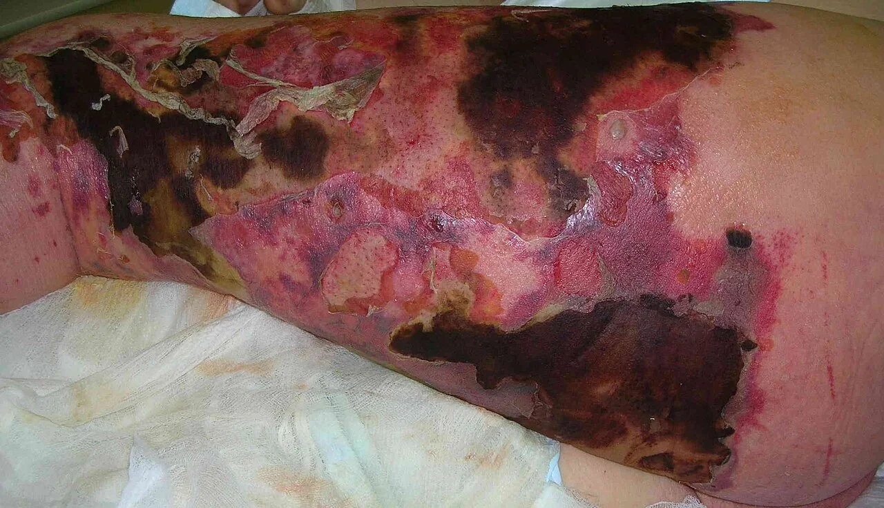
- Severe pain, out of proportion to the visible skin findings - the single most important clue, present in the great majority of cases
- Rapid progression - visible spread over hours rather than days
- Erythema and swelling that may look identical to cellulitis early on
- Skin changes as it progresses - dusky or purple discolouration, blistering, haemorrhagic bullae, and eventually frank necrosis with a black eschar
- Crepitus, if a gas-forming organism is involved - a useful sign when present, but its absence does not exclude the diagnosis
- Systemic toxicity disproportionate to the apparent extent of skin involvement - fever, tachycardia, and progression to septic shock
- A late, ominous sign: reduced pain or anaesthesia over the affected skin, as the infection destroys the cutaneous nerves - a paradoxical improvement in pain that should never be mistaken for genuine recovery
Differential diagnosis
- Cellulitis and erysipelas - the main differential, and the diagnosis most often mistaken for early necrotising fasciitis
- Gas gangrene (clostridial myonecrosis) - deep muscle involvement with prominent gas and profound toxicity, from Clostridium perfringens
- Pyomyositis - deep muscle infection, usually with a more localised presentation
- Deep vein thrombosis - can cause pain and swelling but lacks progressive skin necrosis and systemic toxicity
- Compartment syndrome - severe pain out of proportion is shared, but the context is usually trauma or a fracture rather than spontaneous infection
Investigations
No investigation should delay surgical exploration when clinical suspicion is high. Investigations support the diagnosis and guide resuscitation, but the diagnosis is ultimately made in theatre.
Blood tests
- FBC, CRP, U&Es, LFTs, clotting screen, glucose and lactate
- Blood cultures before antibiotics, without delaying their administration
- Group and save, given the likelihood of significant blood loss at debridement
- Creatine kinase, which can be markedly elevated if muscle is involved
The LRINEC score
The Laboratory Risk Indicator for Necrotising Fasciitis (LRINEC) combines CRP, white cell count, haemoglobin, sodium, creatinine and glucose into a score out of 13, with a score of 6 or more classified as high risk.2
Imaging
CT or MRI can show fascial thickening, fluid tracking along fascial planes, and gas within the soft tissues, and may help delineate the extent of disease or identify a deep collection. In a patient who is clinically stable with genuine diagnostic uncertainty, imaging can be useful - but in a patient with a clear clinical picture, or one who is deteriorating, imaging must never be allowed to delay theatre.
The definitive test: surgical exploration
A bedside or theatre "finger test" - a small incision down to fascia under local anaesthetic - is diagnostic if it reveals grey, necrotic fascia that fails to bleed, a lack of the normal resistance fascia offers to blunt finger dissection, and thin, foul-smelling "dishwater" fluid. Where suspicion is high, this exploration should not be delayed for any other investigation.
Management
Management runs in three parallel strands, none of which should wait for the others: resuscitation, antibiotics, and urgent surgery.
Resuscitation
Treated as sepsis: the Sepsis Six, aggressive IV fluid resuscitation, and early critical care involvement, since these patients frequently progress to septic or toxic shock.
Antibiotics
Broad-spectrum IV antibiotics are started immediately, covering Gram-positive, Gram-negative and anaerobic organisms - a typical UK regimen combines a broad-spectrum beta-lactam (such as piperacillin-tazobactam or a carbapenem) with clindamycin.3
Surgery
Radical surgical debridement of all necrotic tissue is the definitive treatment, performed as an emergency and often needing to be repeated - a planned "second look" procedure at 24-48 hours is standard, since the extent of non-viable tissue is frequently greater than it first appears. Amputation is sometimes necessary to control the source of infection and save the patient's life.
Adjuncts
IV immunoglobulin is used in some cases of streptococcal toxic shock syndrome complicating necrotising fasciitis, on the rationale that it can neutralise circulating superantigens, though the evidence base is less robust than for surgery and antibiotics. Hyperbaric oxygen therapy has been used historically but is not considered standard of care and must never delay debridement. Ongoing input from plastic surgery for later reconstruction, and from critical care throughout, is routine.
Complications
- Septic shock and multi-organ failure
- Limb loss or amputation
- Streptococcal toxic shock syndrome in type II disease
- Major disfigurement requiring extensive reconstructive surgery
- Significant psychological morbidity in survivors
- Death
Prognosis
Mortality remains substantial even with prompt, appropriate treatment, commonly quoted at 20-30% and higher in those who present late, are older, have multiple comorbidities, or develop septic shock. Delay to surgical debridement is the single most consistent predictor of death across published series - which is precisely why the diagnosis must be pursued aggressively on clinical grounds rather than deferred to a confirmatory test.
Survivors frequently require multiple operations, skin grafting or amputation, and face a prolonged recovery, both physical and psychological.
References
- NICE Clinical Knowledge Summaries. Necrotising fasciitis. Available here
- Wong CH, Khin LW, Heng KS et al. The LRINEC (Laboratory Risk Indicator for Necrotizing Fasciitis) score: a tool for distinguishing necrotizing fasciitis from other soft tissue infections. Critical Care Medicine. 2004. Available here
- Stevens DL, Bisno AL, Chambers HF et al. Practice guidelines for the diagnosis and management of skin and soft tissue infections. Clinical Infectious Diseases. 2014. Available here
- Sartelli M, Guirao X, Hardcastle TC et al. 2018 WSES/SIS-E consensus conference: recommendations for the management of skin and soft-tissue infections. World Journal of Emergency Surgery. 2018. Available here
- Faraklas I, Yang H, Kowal-Vern A et al. A multi-center review of necrotizing fasciitis and its management. Journal of Burn Care and Research. 2013.
This article is written for revision and education. It is not clinical guidance and must not be used to make decisions about the care of a patient. Always check current NICE guidance and local protocols.