
Hernias: Groin, Ventral and Incisional
Key points
- Hernia: protrusion of a viscus, or part of one, through a defect in the wall of the cavity that normally contains it.
- The four states: reducible, irreducible (incarcerated), obstructed, and strangulated. Only the last means the blood supply has failed.
- Indirect inguinal: passes through the deep ring, lateral to the inferior epigastric vessels, and can reach the scrotum. The commonest hernia in both sexes.
- Direct inguinal: pushes through a weakness in the posterior wall of the canal, medial to the inferior epigastric vessels, and rarely reaches the scrotum.
- Femoral: below and lateral to the pubic tubercle, commoner in women, and strangulates in up to 40% because the femoral ring is narrow and rigid.
- Surface landmarks: the deep ring lies at the midpoint of the inguinal ligament; the superficial ring lies superomedial to the pubic tubercle.
- Repair: tension-free mesh repair, open (Lichtenstein) or laparoscopic. Laparoscopic repair is preferred for bilateral and recurrent hernias.
- Urgency: femoral hernias and any symptomatic or irreducible hernia should be repaired promptly; asymptomatic inguinal hernias in men may be watched.
Introduction and terminology
A hernia is the protrusion of a viscus, or part of a viscus, through a defect in the wall of the cavity that normally contains it. Around 100,000 groin hernia repairs are performed in the UK each year, making it one of the commonest operations in general surgery.1
Every hernia has three components: the defect in the wall, the sac of peritoneum pushed through it, and the contents of the sac, which may be omentum, small bowel, colon, bladder or, rarely, an ovary. Whether a hernia is dangerous depends far less on its size than on the size and rigidity of the defect - a small, tight neck strangulates, a large lax one does not.
| State | Definition | Clinical picture |
|---|---|---|
| Reducible | Contents return to the abdominal cavity spontaneously or with gentle pressure | A lump that disappears on lying flat and returns on standing or coughing. Usually painless. |
| Irreducible (incarcerated) | Contents cannot be returned, usually because of adhesions within the sac | A persistent lump that does not disappear. Often painless, but strangulation cannot be excluded clinically. |
| Obstructed | The lumen of contained bowel is occluded, but the blood supply is intact | Colicky pain, vomiting, distension and absolute constipation, with a tense hernia |
| Strangulated | The blood supply to the contents is compromised | Constant severe pain, a tense, tender, irreducible, hot lump with overlying erythema, no cough impulse, and systemic signs of sepsis. A surgical emergency. |
The absence of a cough impulse in a previously reducible hernia is a warning sign, not a reassuring one - it means the neck is now occluded.
Anatomy of the inguinal canal
The inguinal canal is an oblique passage about 4 cm long running from the deep to the superficial inguinal ring, carrying the spermatic cord in men and the round ligament in women. It exists because the testis descends through the abdominal wall during development, and its obliquity is what normally keeps it closed.
| Wall | Structures |
|---|---|
| Anterior | Aponeurosis of external oblique along its whole length, reinforced laterally by internal oblique |
| Posterior | Transversalis fascia along its whole length, reinforced medially by the conjoint tendon |
| Roof | Arching fibres of internal oblique and transversus abdominis |
| Floor | Inguinal ligament, reinforced medially by the lacunar ligament |
Two surface landmarks are examined constantly and confused constantly:
- The deep (internal) ring lies at the midpoint of the inguinal ligament, which is halfway between the anterior superior iliac spine and the pubic tubercle. It is a defect in transversalis fascia.
- The superficial (external) ring lies superomedial to the pubic tubercle, and is a triangular defect in the external oblique aponeurosis.
- The mid-inguinal point is a different landmark, halfway between the anterior superior iliac spine and the pubic symphysis, and marks the femoral artery. It is not the deep ring, and examiners test this distinction.
Hesselbach triangle is the area of the posterior wall bounded medially by the lateral border of rectus abdominis, laterally by the inferior epigastric vessels, and inferiorly by the inguinal ligament. A hernia pushing directly through this triangle is a direct hernia; one entering the canal through the deep ring, lateral to the inferior epigastric vessels, is indirect.
The femoral canal is the medial compartment of the femoral sheath, bounded anteriorly by the inguinal ligament, posteriorly by the pectineal (Cooper) ligament, medially by the lacunar ligament and laterally by the femoral vein. It normally contains only fat and a lymph node. It is narrow, and three of its four boundaries are unyielding ligament or bone, which is exactly why a femoral hernia strangulates so readily.
Types of hernia
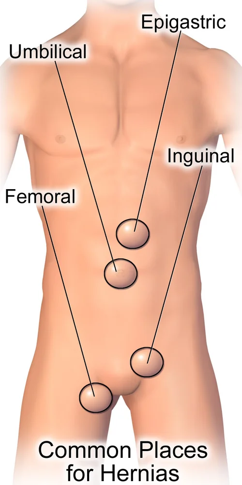
Groin hernias
| Indirect inguinal | Direct inguinal | Femoral | |
|---|---|---|---|
| Route | Through the deep ring, along the canal, out through the superficial ring | Directly through the weakened posterior wall in Hesselbach triangle | Through the femoral canal, below the inguinal ligament |
| Relation to inferior epigastric vessels | Lateral | Medial | Not applicable - below the ligament |
| Relation to pubic tubercle | Above and medial | Above and medial | Below and lateral |
| Cause | Congenital patent processus vaginalis | Acquired muscular weakness | Acquired widening of the femoral ring |
| Typical patient | Any age, including infants and young men | Older men | Older women, especially multiparous |
| Can reach the scrotum | Yes | Rarely | No |
| Controlled by pressure over the deep ring | Yes | No | No |
| Risk of strangulation | Moderate | Low | High - up to 40% |
Around 75% of abdominal wall hernias are inguinal, and about two thirds of those are indirect. Inguinal hernias are far commoner in men, with a lifetime risk of roughly 27% compared with 3% in women. Femoral hernias account for under 5% of groin hernias, but a much larger share of emergency hernia surgery.
Ventral and other hernias
- Umbilical - through the umbilical ring. Common in infants, where the great majority close spontaneously by the age of four or five, so repair is deferred.
- Paraumbilical - through a defect adjacent to the umbilicus in adults, associated with obesity, ascites and multiparity. The neck is often narrow, so these do strangulate and are usually repaired.
- Epigastric - through the linea alba between xiphisternum and umbilicus, usually containing only extraperitoneal fat but often painful
- Incisional - through the scar of a previous laparotomy, occurring in 10 to 20% of midline incisions, with obesity, wound infection, smoking, steroids and poor closure technique as risk factors
- Parastomal - alongside a stoma, extremely common and often managed conservatively
- Spigelian - through the linea semilunaris at the lateral border of rectus, typically below the arcuate line. It lies beneath the intact external oblique aponeurosis, so it is easily missed on examination and often needs imaging.
- Obturator - through the obturator foramen, in thin elderly women. It presents with bowel obstruction and the Howship-Romberg sign, medial thigh pain on internal rotation of the hip from obturator nerve compression.
- Lumbar - through the superior (Grynfeltt) or inferior (Petit) lumbar triangle, rare
Named varieties by contents
- Richter hernia - only part of the circumference of the bowel wall is caught in the sac. It can strangulate and perforate without ever causing obstruction, so the patient has a tender lump and sepsis but is still passing flatus. This is a classic trap.
- Littre hernia - contains a Meckel diverticulum
- Maydl hernia - a W-shaped loop in which the segment lying inside the abdomen between the two limbs is the part that becomes ischaemic, so the bowel within the sac may look healthy
- Sliding hernia - a retroperitoneal organ, commonly caecum, sigmoid or bladder, forms part of the wall of the sac itself rather than lying inside it, which matters at operation because opening the sac risks injuring it
- Amyand hernia - contains the appendix
Risk factors
- Increasing age and male sex, for inguinal hernias
- Chronically raised intra-abdominal pressure - chronic cough, constipation and straining, bladder outflow obstruction, ascites, heavy manual work and obesity
- Connective tissue disorders - Marfan and Ehlers-Danlos syndromes, and abnormalities of collagen metabolism seen in smokers and in patients with abdominal aortic aneurysm
- Previous surgery - for incisional and parastomal hernias, with wound infection the single largest modifiable factor
- Prematurity and low birth weight - for indirect inguinal hernias in infants, through a patent processus vaginalis
- Pregnancy and multiparity - for femoral and umbilical hernias
- Smoking, malnutrition, corticosteroids and diabetes, all of which impair wound healing
Clinical features and examination
Most hernias present as a lump that appears on standing, coughing or straining and disappears on lying down, with a dragging discomfort rather than pain. Pain that is constant and severe is the abnormal finding.
Examining a groin lump
- Examine the patient standing first. A small hernia may be invisible when supine, and examining only on the couch is the commonest reason one is missed.
- Inspect both groins and the scrotum for asymmetry, scars and skin changes, and ask the patient to cough
- Palpate the lump and define its relation to the pubic tubercle - above and medial is inguinal, below and lateral is femoral. Identify the tubercle by tracing the adductor longus tendon upwards.
- Assess for a cough impulse and whether the lump is reducible, ideally by asking the patient to reduce it themselves
- Determine whether you can get above the lump - if not, it is arising from the inguinal canal rather than the scrotum
- Examine the abdomen for distension, tenderness and bowel sounds, and the contralateral side
- Assess for an underlying cause - a chronic cough, prostatic symptoms, constipation, or, in an older patient with a new hernia, an abdominal or pelvic mass
The classical deep ring occlusion test - reducing the hernia, pressing over the deep ring at the midpoint of the inguinal ligament, and asking the patient to cough - is described as distinguishing indirect (controlled) from direct (not controlled) hernias. In practice it is unreliable, and the distinction is made at operation. Say that you would perform it, but do not build your answer on the result.
Differential diagnosis of a groin lump
- Inguinal or femoral hernia, and a saphena varix, which has a fluid thrill on coughing and disappears on lying down
- Inguinal lymphadenopathy - look for a source in the leg, perineum and genitalia
- Femoral artery aneurysm or pseudoaneurysm, which is pulsatile and expansile
- Undescended or ectopic testis, and always confirm both testes are in the scrotum in a boy
- Hydrocele of the cord, epididymal cyst, or a lipoma of the cord
- Psoas abscess, which is fluctuant and associated with spinal pathology
Investigations
Groin hernia is a clinical diagnosis and most need no investigation. Imaging is reserved for genuine uncertainty.
- Ultrasound - first line where a hernia is suspected but not palpable, or to distinguish a hernia from a lymph node, hydrocele or saphena varix. It is dynamic and can be performed with the patient straining.
- CT of the abdomen and pelvis - for suspected obstruction or strangulation, for incisional and Spigelian hernias, and to plan complex repairs by defining the defect and any loss of domain
- MRI - occasionally used for occult groin pain where the differential includes a sportsman groin or hip pathology
- Bloods - FBC, U&Es, CRP, lactate, and group and save where surgery is likely, with a venous gas in any patient with suspected strangulation
Management
Who needs an operation
- All femoral hernias should be repaired, and promptly, because of the high strangulation risk
- All symptomatic hernias, and all irreducible ones
- Paraumbilical, epigastric, Spigelian and obturator hernias, because the necks are narrow
- Asymptomatic or minimally symptomatic inguinal hernias in men may reasonably be watched. Trials of watchful waiting show a low rate of acute events, though most patients eventually cross over to surgery because symptoms develop.2
- Umbilical hernias in children are observed, since most close spontaneously by the age of four or five. Repair is considered if they persist beyond that, or if the defect is large or symptomatic.
- Incisional and parastomal hernias are repaired for symptoms, obstruction or cosmesis, weighed against a high recurrence rate and the difficulty of the operation
Elective repair
The principle is a tension-free repair using a prosthetic mesh, which reduces recurrence several-fold compared with suture repair. The main options are:
| Open (Lichtenstein) | Laparoscopic (TAPP or TEP) | |
|---|---|---|
| Technique | Mesh sutured over the posterior wall of the inguinal canal through a groin incision | Mesh placed in the preperitoneal space, either transabdominally (TAPP) or totally extraperitoneally (TEP) |
| Anaesthesia | Can be done under local or regional anaesthesia, useful in unfit patients | Requires general anaesthesia |
| Recovery | Slightly slower return to work | Less post-operative pain, faster return to normal activity |
| Recurrence | Comparable in experienced hands | Comparable in experienced hands |
| Best suited to | Primary unilateral hernia, patients unfit for general anaesthesia | Bilateral hernias, recurrent hernias after open repair, and patients who need to return to heavy work quickly |
| Drawbacks | Higher rate of chronic wound and nerve pain in some series | Longer learning curve, risk of visceral and vascular injury, higher equipment cost |
NICE recommends that laparoscopic repair be offered as an option for inguinal hernia repair, and it is specifically preferred for bilateral and recurrent hernias.3 Femoral hernias are repaired by closing the femoral canal, and a laparoscopic or open preperitoneal approach handles both inguinal and femoral defects at once, which is one argument for it in women.
Emergency management of a strangulated hernia
- Do not attempt forceful reduction. Reducing dead bowel into the abdomen (reduction en masse) converts a visible problem into a hidden one and can be fatal.
- Resuscitate - intravenous fluids, analgesia, nil by mouth, nasogastric tube if obstructed, catheterise and monitor urine output
- Broad-spectrum intravenous antibiotics and blood cultures if septic
- Urgent senior surgical review and theatre, with group and save or crossmatch
- At operation, the sac is opened, the contents inspected, non-viable bowel resected, and the defect repaired. Mesh is generally avoided where there is frank contamination from perforated bowel, and a suture repair is used instead.
Complications
Of the hernia
- Incarceration, obstruction and strangulation, with bowel infarction, perforation and peritonitis
- Reduction en masse - the hernia and its constricting neck are pushed back together, so the obstruction persists inside the abdomen while the lump has apparently resolved
- Loss of domain in very large longstanding hernias, where returning the contents raises intra-abdominal pressure enough to compromise respiration and renal perfusion
- Skin excoriation and ulceration over large hernias
Of the repair
- Chronic post-herniorrhaphy groin pain, reported by up to 10% of patients and the commonest long-term complication. It arises from injury or entrapment of the ilioinguinal, iliohypogastric or genital branch of the genitofemoral nerve.
- Recurrence, which is low after mesh repair of a primary hernia but substantially higher after incisional and parastomal repairs
- Haematoma and seroma, common and usually self-limiting
- Wound or mesh infection, which may require removal of the mesh
- Ischaemic orchitis and testicular atrophy, from damage to the pampiniform plexus during dissection of the cord
- Injury to the vas deferens, bladder, bowel or femoral vessels
- Urinary retention, particularly after bilateral repair or spinal anaesthesia
Red flags
Prognosis
Elective mesh repair of a primary groin hernia is a highly successful operation. Recurrence rates are in the low single figures, most patients return to normal activity within two to four weeks, and mortality is negligible in fit patients.
Emergency repair is a different operation with different outcomes. Mortality after emergency hernia surgery, particularly where bowel resection is required in an elderly patient, is an order of magnitude higher than after elective repair. This asymmetry is the central argument for repairing femoral hernias promptly and for not allowing a symptomatic inguinal hernia to drift on a long waiting list.
The main long-term issue after successful repair is not recurrence but chronic groin pain, which affects a meaningful minority and can be more disabling than the original hernia. It is one reason why watchful waiting is a legitimate option for a genuinely asymptomatic inguinal hernia in a man, and why the discussion before surgery should include it rather than treating repair as automatic.
References
- HerniaSurge Group. International guidelines for groin hernia management. Hernia. 2018. Available here
- Fitzgibbons RJ, Giobbie-Hurder A, Gibbs JO et al. Watchful waiting vs repair of inguinal hernia in minimally symptomatic men: a randomized clinical trial. JAMA. 2006. Available here
- NICE TA83. Laparoscopic surgery for inguinal hernia repair. 2004. Available here
This article is written for revision and education. It is not clinical guidance and must not be used to make decisions about the care of a patient. Always check current NICE guidance and local protocols.