
Testicular Cancer
Key points
- Testicular cancer: the commonest solid malignancy in men aged 20-40. Over 95% are germ cell tumours, divided into seminomas and non-seminomatous germ cell tumours (NSGCT).
- Classic presentation: a painless, hard, irregular lump arising from the body of the testis that does not transilluminate and that you cannot get above.
- Biggest risk factor: cryptorchidism (undescended testis) - a several-fold increase, with risk in both testes; orchidopexy reduces but does not abolish it.
- Tumour markers: AFP, beta-hCG and LDH - always taken BEFORE orchidectomy. AFP is never raised in a pure seminoma - a raised AFP means the tumour is treated as a non-seminoma.
- First-line imaging: scrotal ultrasound, followed by CT chest, abdomen and pelvis for staging. Spread is to the para-aortic nodes, following embryological drainage.
- Biopsy is contraindicated: never biopsy a suspected testicular tumour through the scrotum - it risks seeding and altering lymphatic drainage. Diagnosis is by inguinal (radical) orchidectomy.
- Fertility: offer sperm banking to every man before treatment, and always before orchidectomy or chemotherapy.
- Prognosis: excellent - over 95% 5-year survival overall, and still around 80% even with metastatic disease. It is one of the most curable solid cancers.
Introduction
Testicular cancer is uncommon overall - around 2,400 UK cases per year, roughly 1% of male cancers - but it is the commonest solid malignancy in men aged 20 to 40, which gives it a clinical importance out of proportion to its incidence.1
Over 95% arise from germ cells, and the fundamental division is between seminomas and non-seminomatous germ cell tumours (NSGCT), which differ in age of presentation, tumour markers, radiosensitivity and treatment. The remainder are non-germ-cell tumours (Leydig and Sertoli cell tumours) or lymphoma, which is the commonest testicular tumour in men over 60.
It is also, importantly, one of the most curable of all solid cancers, with survival exceeding 95% - a transformation driven by cisplatin-based chemotherapy. That makes prompt recognition genuinely worthwhile, and it also means that discussions about fertility preservation and long-term treatment toxicity matter enormously, because most of these men will live for decades afterwards.
| Type | Peak age | Markers | Notes |
|---|---|---|---|
| Seminoma | 30-40 (slightly older) | AFP NEVER raised; hCG raised in ~15%; LDH may be raised | Radiosensitive and very chemosensitive. Grows more slowly. Homogeneous on ultrasound |
| Non-seminomatous (NSGCT) overall | 20-30 (younger) | AFP and/or hCG raised in ~85% | More aggressive, metastasise earlier; treated with chemotherapy rather than radiotherapy |
| - Yolk sac tumour | Children; also adults | AFP markedly raised | Commonest testicular tumour in young boys |
| - Choriocarcinoma | 20-30 | hCG markedly raised | Highly aggressive, haematogenous spread; very high hCG can cause gynaecomastia and thyrotoxicosis |
| - Embryonal carcinoma | 20-30 | AFP and/or hCG | Aggressive; often a component of mixed tumours |
| - Teratoma | 20-30 | AFP variable | In adults may be malignant; mature teratoma is chemoresistant and needs surgical resection |
| Lymphoma | Over 60 | Negative germ cell markers | The commonest testicular malignancy in older men; often bilateral |
Risk factors
- Cryptorchidism (undescended testis) - the strongest risk factor, increasing risk around 3 to 8 fold. Crucially, the risk is raised in both testes, not only the undescended one, implying a shared developmental abnormality. Orchidopexy reduces the risk and, importantly, makes the testis examinable, but does not eliminate it - and the earlier it is performed, the greater the benefit
- Previous testicular cancer - a contralateral tumour develops in around 2-5%, so the remaining testis needs lifelong awareness
- Family history - a father or brother with testicular cancer increases risk several-fold
- Age 20-40 for germ cell tumours
- White ethnicity - incidence is notably higher in white men than in Black or Asian men
- Infertility and poor semen quality - associated independently of cryptorchidism
- Testicular microlithiasis - an ultrasound finding; significant mainly in combination with other risk factors
- Klinefelter syndrome - associated particularly with mediastinal germ cell tumours
- HIV infection - increased risk of seminoma
- Testicular dysgenesis syndrome - the concept linking cryptorchidism, hypospadias, poor semen quality and testicular cancer as manifestations of disordered testicular development
Note what is not a risk factor: trauma and vasectomy do not cause testicular cancer, though trauma often draws attention to a lump that was already present - a useful point when taking a history, since patients frequently attribute the lump to an injury.
Clinical features
Local features
- A painless lump in the testis - the classic presentation, typically noticed by the patient or a partner
- Hard, firm and irregular, arising from the body of the testis itself rather than separate from it
- Does not transilluminate (unlike a hydrocoele)
- You can get above it - it is confined to the scrotum, unlike an inguinoscrotal hernia
- Painless in around 70-80%, but a minority present with dull scrotal ache or heaviness, and around 10% with acute pain from haemorrhage or infarction into the tumour - which can be mistaken for epididymo-orchitis or torsion
- A secondary hydrocoele may be present and can obscure the underlying mass
- A lump that does not resolve after a trial of antibiotics for presumed epididymo-orchitis must be re-examined and scanned - this is a common route to delayed diagnosis
Features of metastatic disease
- Back pain - from para-aortic lymphadenopathy; a classic and easily misattributed symptom in a young man
- Abdominal mass from bulky retroperitoneal nodes
- Cough, haemoptysis or breathlessness from pulmonary metastases
- Supraclavicular lymphadenopathy (Virchow's node)
- Weight loss and malaise
- Gynaecomastia - from beta-hCG secretion, which is structurally similar to LH and stimulates oestrogen production; seen particularly with choriocarcinoma. Very high hCG can also cause thyrotoxicosis through cross-reactivity at the TSH receptor
- Lower limb swelling from venous or lymphatic obstruction
Differential diagnosis
| Condition | Distinguishing features |
|---|---|
| Testicular tumour | Hard, irregular, arising from the testis, does not transilluminate, can get above it |
| Hydrocoele | Fluctuant, transilluminates, testis usually impalpable within it. A new hydrocoele in a young man still warrants ultrasound to exclude an underlying tumour |
| Epididymal cyst / spermatocoele | Separate from and above/behind the testis, smooth, transilluminates |
| Varicocoele | 'Bag of worms', usually left-sided, more prominent on standing and decompresses on lying flat |
| Epididymo-orchitis | Painful, tender, may be febrile, urinary or sexual symptoms, relieved by elevation (positive Prehn's) |
| Testicular torsion | Sudden severe pain, high-riding horizontal testis, absent cremasteric reflex - emergency |
| Inguinoscrotal hernia | Cannot get above it, may be reducible, cough impulse, bowel sounds |
| Testicular lymphoma | Older men (over 60), often bilateral, rapidly enlarging |
| Haematocoele / trauma | History of injury; still requires ultrasound to exclude an underlying tumour |
Investigations
Imaging
- Scrotal ultrasound with Doppler - the first-line investigation, with sensitivity approaching 100% for intratesticular masses. It distinguishes intratesticular (suspicious) from extratesticular (usually benign) lesions
- CT chest, abdomen and pelvis - for staging, assessing the para-aortic nodes, lungs, liver and mediastinum
- MRI or CT head - only if there are neurological symptoms or very high-risk disease (choriocarcinoma has a particular propensity for brain metastases)
- Chest X-ray - may show pulmonary metastases, but CT is required for staging
Tumour markers - taken BEFORE orchidectomy
- Alpha-fetoprotein (AFP) - never raised in pure seminoma. Raised in yolk sac tumours and many embryonal carcinomas. Half-life 5-7 days
- Beta-human chorionic gonadotrophin (beta-hCG) - raised in choriocarcinoma (often markedly), embryonal carcinoma, and around 15% of seminomas. Half-life 24-36 hours
- Lactate dehydrogenase (LDH) - a non-specific marker of tumour bulk and cell turnover; useful prognostically
- Why before surgery: pre-operative levels are needed for staging, risk stratification (the IGCCCG classification) and to interpret post-operative decline. Markers should fall according to their half-lives after orchidectomy; failure to normalise indicates residual disease
- Markers are also used for surveillance and to detect relapse, often before it is radiologically apparent
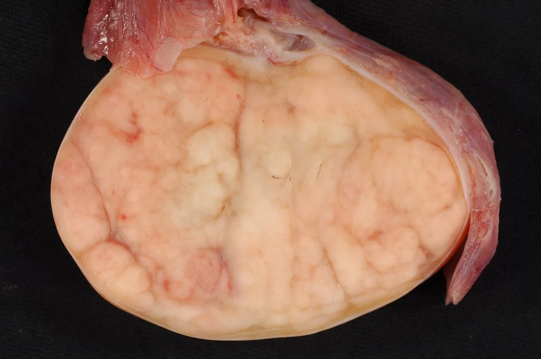
Tissue diagnosis
Staging
| Stage | Description |
|---|---|
| I | Confined to the testis, no evidence of metastasis |
| II | Infradiaphragmatic nodal involvement (para-aortic nodes), subdivided by node size: IIa <2 cm, IIb 2-5 cm, IIc >5 cm |
| III | Supradiaphragmatic nodal involvement |
| IV | Extralymphatic metastases - lung, liver, bone, brain |
Metastatic disease is further stratified into good, intermediate and poor prognosis groups by the International Germ Cell Cancer Collaborative Group (IGCCCG) classification, using marker levels, primary site and the presence of non-pulmonary visceral metastases. This directly determines the number of chemotherapy cycles.
Management
Before any treatment
Surgery
Radical inguinal orchidectomy is both diagnostic and therapeutic, and is performed in essentially all cases. Retroperitoneal lymph node dissection (RPLND) may be used in selected NSGCT patients, particularly for residual masses after chemotherapy, since mature teratoma within a residual mass is chemoresistant and must be resected.
Adjuvant and systemic treatment
| Situation | Management |
|---|---|
| Stage I seminoma | Surveillance is standard for most (relapse ~15-20%, and salvage is highly effective). Alternatives are a single dose of carboplatin or para-aortic radiotherapy in selected patients |
| Stage I NSGCT | Surveillance, or one cycle of BEP chemotherapy if high risk (notably lymphovascular invasion) |
| Metastatic seminoma or NSGCT | BEP chemotherapy - Bleomycin, Etoposide, cisPlatin - typically 3-4 cycles depending on IGCCCG risk group |
| Residual mass after chemotherapy | Seminoma: often observed or assessed with PET-CT. NSGCT: surgical resection (RPLND), because residual mature teratoma is chemoresistant and can grow or transform |
| Relapsed disease | Salvage chemotherapy, sometimes with high-dose chemotherapy and stem cell rescue - still with meaningful cure rates |
Complications
- Metastatic disease - para-aortic nodes, lung, liver, brain; choriocarcinoma in particular spreads early and haematogenously
- Infertility and subfertility - from the disease itself, orchidectomy, chemotherapy and radiotherapy
- Hypogonadism - requiring testosterone replacement, particularly after bilateral disease
- Contralateral testicular tumour - in 2-5% of survivors
- Bleomycin-induced pulmonary fibrosis - and the lifelong risk associated with high-concentration oxygen
- Cisplatin toxicity - chronic kidney disease, permanent hearing loss and tinnitus, peripheral neuropathy, and Raynaud phenomenon
- Secondary malignancy - leukaemia from etoposide, and solid tumours after radiotherapy
- Cardiovascular disease and metabolic syndrome - significantly increased in long-term survivors
- Retrograde ejaculation after retroperitoneal lymph node dissection, from damage to sympathetic nerves (nerve-sparing techniques reduce this)
- Psychological effects - body image, fertility concerns, and anxiety about recurrence
- Tumour lysis syndrome - rare, with very bulky disease starting chemotherapy
Red flags
Prognosis
Testicular cancer has among the best outcomes of any solid malignancy. Overall 5-year survival in the UK exceeds 95%, and for stage I disease it approaches 99%. Even metastatic disease is frequently curable, with around 80% long-term survival overall and over 90% in the good-prognosis IGCCCG group. This is one of the great successes of modern oncology, and it rests almost entirely on the introduction of cisplatin-based combination chemotherapy in the 1970s, which converted a usually fatal disease into a usually curable one.
Prognosis is determined by stage, histological type, marker levels and the IGCCCG risk group. Seminomas generally do better than non-seminomatous tumours, being both radiosensitive and highly chemosensitive, and tending to present earlier and spread in a more predictable, orderly fashion. Adverse features include very high markers, non-pulmonary visceral metastases (liver, bone, brain) and a mediastinal primary.
Because survival is so good and patients are so young, the dominant clinical concern shifts to survivorship. Men cured of testicular cancer face decades of potential late effects: cardiovascular disease and metabolic syndrome, hypogonadism, cisplatin-related renal impairment, hearing loss and neuropathy, bleomycin-related pulmonary fibrosis, second malignancies, and subfertility. There is also a 2-5% risk of a contralateral tumour, which is why these men should be taught testicular self-examination and followed up long term. Counselling at diagnosis should therefore convey genuine optimism about cure while being honest that treatment carries consequences which will need managing for the rest of their lives.
References
- NICE NG12. Suspected cancer: recognition and referral. 2015, updated 2023. Available here
- Cancer Research UK. Testicular cancer statistics. Available here
- European Association of Urology. Guidelines on Testicular Cancer. Available here
- International Germ Cell Cancer Collaborative Group. IGCCCG prognostic factor-based staging system. Journal of Clinical Oncology. 1997. Available here
- NICE Clinical Knowledge Summaries. Scrotal pain and swelling. Available here
- BNF. Bleomycin - cautions and pulmonary toxicity. Available here
- Ed Uthman, MD, CC BY 2.0, via Wikimedia Commons. Available here
This article is written for revision and education. It is not clinical guidance and must not be used to make decisions about the care of a patient. Always check current NICE guidance and local protocols.