
Benign Eyelid Disorders
Key points
- Blepharitis: chronic inflammation of the lid margins, anterior (staphylococcal or seborrhoeic) or posterior (meibomian gland dysfunction). Managed, not cured, with lid hygiene.
- Stye (hordeolum): an acute, tender, localised infection of a lash follicle (external) or meibomian gland (internal). Warm compresses; antibiotics only if there is surrounding cellulitis.
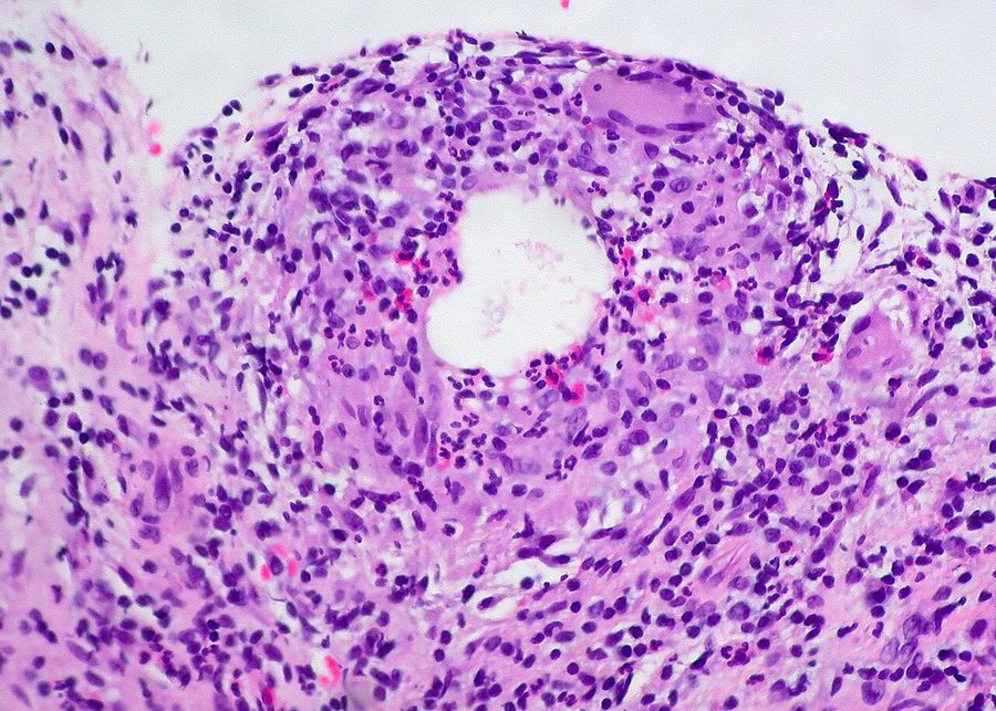
- Chalazion: a painless, firm, rubbery granuloma of a blocked meibomian gland. Warm compresses and massage; incision and curettage if it persists beyond 4-6 weeks.
- Entropion: the lid turns inward, so lashes abrade the cornea. Taping and lubricants as a holding measure; definitive treatment is surgical.
- Ectropion: the lid turns outward, causing watering, exposure keratopathy and a chronically red eye. Lubricants and surgery.
- Ptosis: a droopy upper lid. The critical distinction is between benign aponeurotic ptosis and the sinister causes - third nerve palsy and Horner syndrome.
- Red flag lid lesion: loss of lashes (madarosis), ulceration, induration, telangiectasia, distortion of the lid margin and recurrence after excision suggest malignancy.
- The one to know: a recurrent unilateral chalazion, particularly in an older patient, may be a sebaceous gland carcinoma masquerading as a benign lump - biopsy it.
Introduction
Eyelid disorders are among the commonest reasons people present with an eye complaint, and the great majority are benign, chronic and manageable in primary care. They are also, taken collectively, a substantial source of misery: chronic blepharitis and dry eye account for an enormous number of consultations, and patients are frequently dissatisfied because they have been told the problem is minor without being told that it is also permanent.
The eyelids do more than close. They spread the tear film with every blink, contribute the lipid layer that stops it evaporating, pump tears through the lacrimal drainage system, and protect the globe mechanically. Disorders of lid position, lid margin health or lid closure therefore produce ocular surface disease, and this is why an apparently trivial lid problem can present as a chronically red, gritty, watering eye.
This article covers the benign conditions and, importantly, the features that should make you doubt that a lid lesion is benign at all. Basal cell carcinoma is the commonest periocular malignancy, and the majority of lid cancers present as something the patient - and sometimes the clinician - assumed was a cyst.
Relevant anatomy
- Skin and orbicularis oculi - the thinnest skin in the body, over the sphincter muscle supplied by the facial nerve, which closes the eye
- Tarsal plate - the dense fibrous skeleton of the lid, containing the meibomian glands which secrete the lipid layer of the tear film through orifices along the posterior lid margin
- Levator palpebrae superioris - opens the upper lid, supplied by the oculomotor nerve, inserting via an aponeurosis into the tarsus and skin
- Müller's muscle - a small smooth muscle contributing 2 mm of lid elevation, under sympathetic control. Its loss explains the partial ptosis of Horner syndrome.
- Glands of Zeis and Moll - sebaceous and apocrine glands associated with the lash follicles at the anterior lid margin
- Grey line - the surface marking dividing anterior (skin and orbicularis) from posterior (tarsus and conjunctiva) lamellae, and the surgical landmark for lid splitting
- Puncta and canaliculi - the drainage openings at the medial end of each lid, requiring correct lid apposition to work
Blepharitis
Chronic inflammation of the lid margins, and one of the commonest conditions in ophthalmology. It is bilateral, fluctuating, and characteristically worse in the morning.
| Feature | Anterior blepharitis | Posterior blepharitis (MGD) |
|---|---|---|
| Site | Lash line and skin of the lid margin | Meibomian gland orifices along the posterior margin |
| Cause | Staphylococcus colonisation, or seborrhoeic dermatitis | Meibomian gland dysfunction with thickened, stagnant secretions |
| Signs | Crusting, collarettes around lash bases, lash loss, ulceration at the margin in staphylococcal disease; greasy scales in seborrhoeic disease | Capped, plugged or pouting gland orifices, thickened and irregular lid margin, frothy or oily tear film, telangiectasia |
| Associations | Seborrhoeic dermatitis, atopic eczema, Demodex infestation | Acne rosacea, dry eye disease |
| Complications | Stye, marginal keratitis, trichiasis, madarosis, corneal punctate erosions | Chalazion, evaporative dry eye, recurrent corneal erosion, chalazia in crops |
Management
- Warm compresses - a heated eye bag or flannel applied for 5-10 minutes, to melt inspissated meibomian secretions
- Lid massage - rolling a finger from the base towards the lid margin to express the softened secretions
- Lid hygiene - cleaning the lid margins with a cotton bud and dilute baby shampoo or a proprietary lid cleanser, once or twice daily
- Preservative-free artificial tears for the associated dry eye
- Topical antibiotic ointment - chloramphenicol rubbed into the lid margin at night for a few weeks, for staphylococcal blepharitis not responding to hygiene alone5
- Oral doxycycline 100 mg daily for 6-12 weeks - for severe meibomian gland dysfunction and rosacea, acting through anti-inflammatory and anti-lipase effects rather than as an antibiotic. Contraindicated in pregnancy, breastfeeding and children under 12, where erythromycin is used instead.
- Treat associated rosacea and seborrhoeic dermatitis
- Consider Demodex where there are cylindrical dandruff-like sleeves around the lashes; treated with tea tree oil preparations
Stye and chalazion
These are frequently confused, and the distinction is simply whether the lesion is acutely infected and painful or a chronic sterile granuloma.
| Feature | Stye (hordeolum) | Chalazion (meibomian cyst) |
|---|---|---|
| Nature | Acute bacterial infection, usually Staphylococcus aureus | Sterile lipogranulomatous inflammation from a blocked meibomian gland |
| Pain | Tender and painful | Painless or only mildly uncomfortable |
| Site | External: at the lash line, pointing forward. Internal: within the tarsus, pointing to the conjunctival surface. | Within the tarsal plate, away from the lid margin |
| Appearance | Red, swollen, with a visible pustular point | Firm, rubbery, discrete, non-tender lump; overlying skin normal |
| Onset | Days | Weeks |
| Treatment | Warm compresses; epilate the lash of an external stye; antibiotic only if surrounding cellulitis | Warm compresses and massage; incision and curettage if persisting beyond 4-6 weeks |
| Course | Discharges and resolves within a week or two | May persist for months; around a third resolve spontaneously |
Malpositions of the lid
Entropion
The lid margin turns inward, so the lashes rub against the cornea and conjunctiva. It is almost always the lower lid, and almost always involutional - the result of age-related horizontal lid laxity, disinsertion of the lower lid retractors and overriding of the preseptal orbicularis.
- Symptoms - a chronically red, gritty, watering, painful eye, often labelled as conjunctivitis for months
- Signs - inturned lid margin and lashes, corneal punctate staining, and in severe cases corneal ulceration and scarring
- Other causes - cicatricial entropion from posterior lamellar scarring in trachoma, Stevens-Johnson syndrome, ocular cicatricial pemphigoid and chemical injury; spastic entropion after ocular surgery or irritation; and congenital entropion
- Temporary measures - lubricating ointment, taping the lower lid down to the cheek, and botulinum toxin to the orbicularis
- Definitive treatment is surgical - a Quickert or Jones procedure to tighten the lid horizontally and reattach the retractors4
- Trichiasis, where the lashes are misdirected but the lid position is normal, is managed by epilation, electrolysis or cryotherapy rather than lid surgery
Ectropion
The lid margin turns outward, away from the globe. Again usually the lower lid and usually involutional.
- Symptoms - epiphora (watering), because the punctum is no longer apposed to the tear lake; a red, sore eye from exposure
- Signs - everted lid margin, exposed palpebral conjunctiva which becomes keratinised and thickened, punctate corneal staining inferiorly, and in severe cases exposure keratopathy
- Causes - involutional (commonest); paralytic in facial nerve palsy; cicatricial from anterior lamellar shortening after burns, sun damage or excessive skin excision; and mechanical from a lid tumour dragging the lid down
- Management - lubricants and ointment at night, taping, and treating any underlying cause. Surgery tightens the lid horizontally, and cicatricial cases need a skin graft to lengthen the anterior lamella.
- Facial nerve palsy deserves particular attention because the combination of ectropion, lagophthalmos and reduced blink threatens the cornea - see the red flag below
Ptosis
Drooping of the upper lid. The clinical task is to separate the common benign cause from the neurological ones, which is done largely on the associated signs - the pupil, the eye movements and the fatigability.
| Cause | Mechanism | Key features |
|---|---|---|
| Aponeurotic (involutional) | Stretching or disinsertion of the levator aponeurosis | By far the commonest; bilateral but asymmetrical, elderly, high lid crease, good levator function, normal pupil and movements. Also after contact lens wear and eye surgery. |
| Third nerve palsy | Paralysis of levator palpebrae superioris | Complete ptosis, eye 'down and out', diplopia. A dilated pupil indicates a compressive lesion - posterior communicating artery aneurysm until proved otherwise. |
| Horner syndrome | Loss of sympathetic supply to Müller's muscle | Partial ptosis (about 2 mm), miosis and anhidrosis, with apparent enophthalmos. Causes include Pancoast tumour, carotid dissection, brainstem stroke and neck lesions. |
| Myasthenia gravis | Neuromuscular junction failure | Fatigable, variable, worse at the end of the day; often with diplopia and Cogan's lid twitch. Ice pack test and acetylcholine receptor antibodies. |
| Myogenic | Muscle disease | Myotonic dystrophy, chronic progressive external ophthalmoplegia, oculopharyngeal muscular dystrophy - bilateral, symmetrical, slowly progressive |
| Mechanical | Weight or scarring | Lid tumour, chalazion, oedema, dermatochalasis, scarring |
| Congenital | Dystrophic levator muscle | Present from birth, poor levator function, lid lag on downgaze. Requires assessment for amblyopia if the pupil is covered. |
Assessment involves measuring the margin-reflex distance (from the corneal light reflex to the upper lid margin, normally 4-5 mm), the palpebral aperture, the levator function (lid excursion from full downgaze to full upgaze with the brow held, normally over 15 mm) and the skin crease position. A high or absent crease points to aponeurotic dehiscence; good levator function with a high crease is the classic involutional picture.
Lid lumps: benign and malignant
Common benign lesions
- Chalazion and stye - as above
- Cyst of Moll - a small, clear, fluid-filled cyst at the lid margin arising from an apocrine gland
- Cyst of Zeis - similar but opaque and yellowish, arising from a sebaceous gland
- Epidermoid (sebaceous) cyst - a mobile, skin-coloured cyst with a central punctum
- Xanthelasma - soft yellow plaques at the medial canthus, associated with dyslipidaemia in around half of cases; check a lipid profile
- Molluscum contagiosum - umbilicated pearly papules; if on the lid margin they cause a chronic follicular conjunctivitis
- Viral wart and papilloma - pedunculated or sessile keratinised lesions
- Naevus - pigmented, usually stable over years
- Dermatochalasis - redundant upper lid skin in older adults, causing a mechanical ptosis and superior field loss
Malignant lesions
- Basal cell carcinoma - around 90% of eyelid malignancies. A pearly, rolled-edge nodule with surface telangiectasia, often centrally ulcerated (rodent ulcer), most commonly on the lower lid and medial canthus. Locally invasive but does not metastasise; medial canthal lesions can invade the orbit and are the most difficult to manage.
- Squamous cell carcinoma - less common, faster growing, hyperkeratotic and ulcerated, with a real risk of perineural spread and nodal metastasis
- Sebaceous gland carcinoma - the masquerader described above, presenting as recurrent chalazion or unilateral chronic blepharitis
- Melanoma - rare on the lid but should be considered for any changing pigmented lesion, applying the standard ABCDE criteria
- Merkel cell carcinoma and Kaposi sarcoma - rare, the latter in advanced HIV
Suspicious lesions are referred for incisional or excisional biopsy, and confirmed malignancies are excised with margin control - Mohs micrographic surgery or frozen section control is preferred periocularly, where tissue conservation matters enormously for lid function.3 Reconstruction depends on the size of the defect and on whether one or both lamellae have been lost.
Watering eye and prognosis
Epiphora is one of the commonest lid-related complaints and has a simple logic: tears either overflow because too many are produced, or because they are not drained.
- Overproduction - paradoxically, dry eye is a common cause, since surface irritation triggers reflex lacrimation. So are blepharitis, trichiasis, entropion and a foreign body.
- Drainage failure - lid position - ectropion or lower lid laxity, so the punctum is not in contact with the tear lake
- Drainage failure - pump failure - orbicularis weakness in facial nerve palsy
- Drainage failure - obstruction - punctal stenosis, canalicular obstruction, or nasolacrimal duct obstruction. In infants this is congenital and resolves spontaneously in around 90% by one year with massage alone.
- Dacryocystitis - infection of an obstructed lacrimal sac, giving a tender red swelling below the medial canthal tendon, treated with oral antibiotics and later with dacryocystorhinostomy
- Investigation - fluorescein dye disappearance test, syringing and probing of the canaliculi, and dacryocystography where indicated
The prognosis for benign lid disease is generally excellent, but with an important qualification about expectations. Blepharitis and meibomian gland dysfunction are chronic relapsing conditions controlled rather than cured, and patients do far better when this is said clearly. Chalazia resolve spontaneously in about a third of cases and respond well to incision and curettage otherwise. Entropion, ectropion and ptosis are corrected reliably by surgery, though involutional problems can recur as tissues continue to age.
The outcome that depends most on the clinician is the recognition of malignancy. Periocular basal cell carcinoma detected early is cured by local excision with an excellent cosmetic result; the same tumour left for two years while treated as a cyst may require orbital exenteration. Applying the madarosis-ulceration-telangiectasia-induration checklist to every lid lump, and biopsying every recurrent chalazion, costs very little and is where the real value of this topic lies.1,2
References
- NICE Clinical Knowledge Summaries. Blepharitis. Available here
- NICE Clinical Knowledge Summaries. Meibomian cyst (chalazion). Available here
- NICE NG12. Suspected cancer: recognition and referral. 2015, updated 2023. Available here
- Royal College of Ophthalmologists. Oculoplastic clinical guidance. Available here
- BNF. Chloramphenicol - eye preparations. Available here
This article is written for revision and education. It is not clinical guidance and must not be used to make decisions about the care of a patient. Always check current NICE guidance and local protocols.