
Blood Group Incompatibility in Pregnancy
Key points
- Mechanism: fetal red cells entering the circulation of an antigen-negative mother provoke maternal IgG, which crosses the placenta and haemolyses fetal red cells.
- Timing: the first sensitising pregnancy is usually unaffected because the primary response is IgM; subsequent pregnancies are affected and worsen progressively.
- Prevalence: around 15% of the UK population is RhD negative; anti-D prophylaxis has reduced deaths from rhesus disease by more than 95%.
- Other antibodies: anti-c and anti-K are the other clinically important ones; anti-K also suppresses fetal erythropoiesis so causes severe anaemia with little jaundice.
- Routine prophylaxis: 1500 IU anti-D at 28 weeks, or 500 IU at 28 and 34 weeks, plus a dose after any potentially sensitising event.
- Kleihauer: performed after 20 weeks to quantify fetomaternal haemorrhage and calculate whether additional anti-D is needed.
- Monitoring: middle cerebral artery peak systolic velocity above 1.5 multiples of the median indicates significant fetal anaemia.
- Treatment: intrauterine transfusion for established fetal anaemia; phototherapy, exchange transfusion and top-up transfusion after birth.
Introduction
Haemolytic disease of the fetus and newborn arises when a mother makes IgG antibodies against an antigen present on her fetus's red cells but absent from her own. Those antibodies cross the placenta, bind fetal red cells, and cause their destruction by splenic macrophages. The consequences range from mild neonatal jaundice to fetal hydrops and intrauterine death.
Rhesus D is by far the most important antigen involved, and its story is one of the genuine triumphs of twentieth-century obstetrics. Before anti-D prophylaxis was introduced in 1969, rhesus disease caused around 46 deaths per 100,000 births in the UK; it now causes fewer than 2. The residual cases arise from failures of the prophylaxis programme rather than from any deficiency in the drug, which is why the sensitising events and the doses are worth learning properly.1,2
Pathophysiology
The RhD antigen is a transmembrane protein encoded by the RHD gene on chromosome 1. Around 15% of the UK population lacks the gene entirely and is RhD negative; the proportion is lower in South Asian and African populations. An RhD negative woman carrying an RhD positive fetus is at risk of sensitisation whenever fetal red cells enter her circulation.
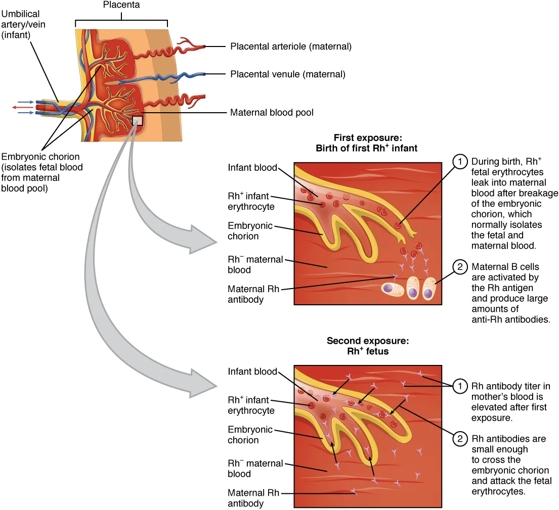
The primary immune response produces IgM, which is a large pentamer and does not cross the placenta, so the sensitising pregnancy itself is almost always unaffected. On re-exposure in a subsequent pregnancy, a brisk secondary response produces IgG, which crosses the placenta by active FcRn-mediated transport and coats fetal red cells. The coated cells are removed by the fetal spleen.
The fetus responds to the resulting anaemia by increasing erythropoiesis, initially in the marrow and then at extramedullary sites, which is why the condition was historically called erythroblastosis fetalis: nucleated red cell precursors appear in the fetal circulation. Hepatic erythropoiesis displaces normal hepatic architecture, portal hypertension develops and albumin synthesis falls. The combination of high-output cardiac failure from severe anaemia and reduced oncotic pressure produces hydrops fetalis, with ascites, pleural and pericardial effusions, skin oedema and placentomegaly.
Which antibodies matter
| Antibody | Significance |
|---|---|
| Anti-D | The commonest cause of severe disease and the only one for which prophylaxis exists |
| Anti-c | The second most important; can cause severe fetal anaemia and hydrops |
| Anti-K (Kell) | Causes severe fetal anaemia by suppressing erythroid precursors as well as by haemolysis; titres correlate poorly with severity and there is relatively little jaundice |
| Anti-E, anti-C, anti-e | Usually mild, but anti-E potentiates anti-c when both are present |
| Anti-Fya (Duffy), anti-Jka (Kidd) | Occasionally cause moderate disease |
| ABO incompatibility | Common, usually mild; a group O mother with IgG anti-A or anti-B and a group A or B infant; can affect a first pregnancy and typically causes neonatal jaundice rather than fetal anaemia |
| Anti-Lewis, anti-P1, anti-M (usually) | Clinically insignificant, being IgM and not crossing the placenta |
Potentially sensitising events
Anti-D immunoglobulin must be given within 72 hours of any event that may have caused a fetomaternal haemorrhage. If it has been missed, it is still worth giving up to 10 days afterwards, since some protection remains.
- Delivery of an RhD positive infant, whether vaginal or by caesarean section
- Miscarriage after 12 weeks, and any miscarriage managed surgically at any gestation
- Threatened miscarriage after 12 weeks, and repeated or heavy bleeding with pain before 12 weeks
- Ectopic pregnancy and molar pregnancy
- Termination of pregnancy at any gestation
- Invasive prenatal testing: amniocentesis, chorionic villus sampling, fetal blood sampling
- Antepartum haemorrhage
- External cephalic version, and any attempt at it
- Abdominal trauma, including road traffic collisions
- Intrauterine death and stillbirth
| Situation | Dose | Kleihauer required? |
|---|---|---|
| Sensitising event before 20 weeks | 250 IU | No |
| Sensitising event at or after 20 weeks | 500 IU | Yes, to determine whether additional doses are needed |
| Routine antenatal prophylaxis | 1500 IU at 28 weeks, or 500 IU at 28 and 34 weeks | No |
| After birth of an RhD positive infant | At least 500 IU within 72 hours | Yes, with 125 IU extra for each additional mL of fetal cells above 4 mL |
Testing
Booking and routine antenatal testing
Every woman has her ABO and RhD group determined and an antibody screen performed at booking, and the antibody screen is repeated at 28 weeks. Where clinically significant antibodies are detected, they are quantified or titred and the woman is referred for specialist assessment.
Cell-free fetal DNA RHD genotyping
Around 40% of RhD negative women in the UK carry an RhD negative fetus and derive no benefit from anti-D. Non-invasive fetal RHD genotyping on maternal plasma from around 11-16 weeks identifies these pregnancies with high accuracy, allowing prophylaxis to be withheld safely. It is now offered by many UK trusts and reduces exposure to a pooled human blood product.2
The Kleihauer test
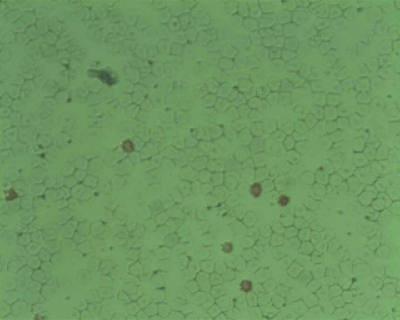
The Kleihauer-Betke acid elution test exploits the resistance of fetal haemoglobin to acid. A maternal blood film is exposed to acid buffer, which elutes adult haemoglobin and leaves maternal cells as pale ghosts, while fetal cells retain their haemoglobin and stain pink. Counting them gives an estimate of the volume of fetal blood in the maternal circulation and hence the anti-D dose required. Flow cytometry is used in some laboratories and is more precise.
Middle cerebral artery Doppler
In a sensitised pregnancy the key question is whether the fetus is anaemic. Anaemic fetal blood has a lower viscosity and the anaemic fetus has a hyperdynamic circulation, so blood flows faster; measuring the peak systolic velocity in the middle cerebral artery therefore detects anaemia non-invasively. A value above 1.5 multiples of the median for gestation indicates moderate to severe fetal anaemia and prompts fetal blood sampling with a view to transfusion.
This has replaced serial amniocentesis for bilirubin measurement using the Liley and Queenan charts, which required repeated invasive procedures that themselves risked further sensitisation. Knowing that the old method existed is useful for interpreting older literature, but MCA Doppler is now the standard of care.1,4
Management of the sensitised pregnancy
- Quantify the antibody at booking and repeat 4-weekly to 28 weeks, then 2-weekly, or more frequently if levels are rising
- Determine the paternal genotype where paternity is certain, or use cell-free fetal DNA genotyping, to establish whether the fetus carries the antigen at all
- Refer to a fetal medicine unit when anti-D exceeds 4 IU/mL, when anti-c exceeds 7.5 IU/mL, when anti-K is detected at any significant titre, or when there is a history of a previously affected pregnancy
- Begin serial middle cerebral artery Doppler surveillance from around 18 weeks in at-risk pregnancies
- Look for early signs of hydrops on ultrasound: ascites, pericardial effusion, skin oedema, placentomegaly and polyhydramnios
- Give antenatal corticosteroids where preterm birth is anticipated
Intrauterine transfusion
Where fetal anaemia is confirmed, packed red cells that are group O, RhD negative, antigen-negative for the relevant antibody, cytomegalovirus negative and irradiated are transfused into the umbilical vein under ultrasound guidance, usually at the cord insertion. Transfusions can be started from around 18 weeks and are repeated at intervals of 2-4 weeks until birth. Survival rates in specialist centres now exceed 90% for non-hydropic fetuses, and remain good even where hydrops has developed. Where disease is very early and severe, maternal intravenous immunoglobulin or plasma exchange may be used to buy time until transfusion becomes technically feasible.1
Timing of birth
Birth is generally planned between 34 and 37 weeks depending on severity and on how many transfusions have been required, balancing the risks of further intrauterine procedures against those of prematurity. The neonatal team must be informed in advance, and antigen-negative crossmatched blood should be available in the delivery unit.
The affected newborn
- Cord bloods at birth for haemoglobin, bilirubin, blood group and a direct antiglobulin (Coombs) test, which is positive in immune haemolysis
- Jaundice within the first 24 hours is always pathological; measure serum bilirubin urgently and plot on the gestation-specific treatment threshold chart
- Phototherapy converts unconjugated bilirubin to water-soluble isomers excreted without conjugation, and is first-line treatment
- Exchange transfusion for bilirubin above the exchange threshold or for severe anaemia, removing both bilirubin and antibody-coated cells
- Intravenous immunoglobulin may reduce the need for exchange transfusion in some cases
- Late anaemia at 4-6 weeks is common, because maternal antibody persists and continues to suppress and destroy red cells long after the jaundice has resolved; arrange follow-up haemoglobin checks
- Kernicterus is the feared complication of untreated hyperbilirubinaemia, causing athetoid cerebral palsy, sensorineural deafness and gaze palsy
Red flags
Prognosis
For an unsensitised RhD negative woman receiving appropriate prophylaxis, the risk of sensitisation across a pregnancy is now well under 1%, and rhesus disease has become rare enough that many clinicians will never see a hydropic fetus. The programme depends entirely on the discipline of giving anti-D correctly and on time, which is why the sensitising events list is examined so persistently.
Once a woman is sensitised, she is sensitised for life, and every subsequent antigen-positive pregnancy will be affected and typically more severely than the last. With modern fetal medicine care, however, outcomes are good: survival after intrauterine transfusion exceeds 90%, and long-term neurodevelopmental outcomes in survivors are largely normal where hydrops did not develop. Women should be counselled before their next pregnancy and booked early with a fetal medicine unit.1,4
References
- BSH Guideline. The management of women with red cell antibodies during pregnancy. British Journal of Haematology. Available here
- RCOG Green-top Guideline No. 22. The use of anti-D immunoglobulin for rhesus D prophylaxis. Available here
- NICE CG98. Jaundice in newborn babies under 28 days. Available here
- Mari G, Deter RL, Carpenter RL et al. Noninvasive diagnosis by Doppler ultrasonography of fetal anemia due to maternal red-cell alloimmunization. N Engl J Med. 2000. Available here
This article is written for revision and education. It is not clinical guidance and must not be used to make decisions about the care of a patient. Always check current NICE guidance and local protocols.