
Osteomyelitis and Septic Arthritis in Practice
Key points
- Routes of infection: haematogenous seeding (commonest in children and the elderly), direct inoculation (trauma, surgery, injection), and spread from a contiguous focus of infection.
- Organism by context: Staphylococcus aureus is the commonest cause overall; think Salmonella in sickle cell disease, Kingella kingae in young children, Neisseria gonorrhoeae in sexually active young adults, and Pseudomonas after a puncture wound through a trainer sole.
- Diagnosis: a hot, swollen, immobile joint is septic arthritis until proven otherwise - aspirate before starting antibiotics whenever the patient is stable enough to allow it.
- In children: the Kocher criteria (non-weight bearing, fever >38.5°C, ESR >40, WCC >12) help separate septic arthritis from transient synovitis, but clinical judgement and joint aspiration remain central.
- Imaging: plain X-ray is often normal for the first 1-2 weeks; MRI is the most sensitive test and the investigation of choice when osteomyelitis is suspected.
- Septic arthritis: needs surgical washout as well as antibiotics - it is a joint emergency, not solely a medical one.
- OVIVA: the 2019 OVIVA trial showed oral antibiotics are non-inferior to IV therapy for bone and joint infection once the patient is stable, provided a highly bioavailable oral agent is used - this changed UK practice away from routine prolonged IV courses.
Introduction
Osteomyelitis is infection of bone; septic arthritis is infection within a joint space. They are considered together here because they share much of their microbiology, their route of spread, and their overall management logic - and because in children the two are anatomically linked, since infection can cross from the metaphysis into the joint through transphyseal vessels that persist until around 18 months of age.
This article focuses on the infectious disease reasoning that runs across both conditions: which organism to suspect in which patient, how to get a microbiological diagnosis before treatment clouds the picture, and how antibiotic strategy has changed in recent years. The orthopaedic detail of surgical washout and fixation is covered in the musculoskeletal notes on osteomyelitis and septic arthritis; this article is the infection-focused companion to those.
Pathophysiology and routes of infection
- Haematogenous spread - bacteraemia seeding bone or a joint; the commonest route in children, where the vascular, looping metaphyseal vessels of growing long bones favour bacterial deposition, and in older or immunosuppressed adults
- Direct inoculation - trauma, an open fracture, surgery, a prosthetic joint, or intra-articular injection introducing organisms directly
- Contiguous spread - infection tracking from an adjacent focus, such as a diabetic foot ulcer extending into the underlying bone, or vertebral osteomyelitis spreading from an epidural or psoas abscess
- Direct extension from metaphysis to joint in young children - via transphyseal vessels present before the growth plate matures, meaning osteomyelitis and septic arthritis can occur together in this age group
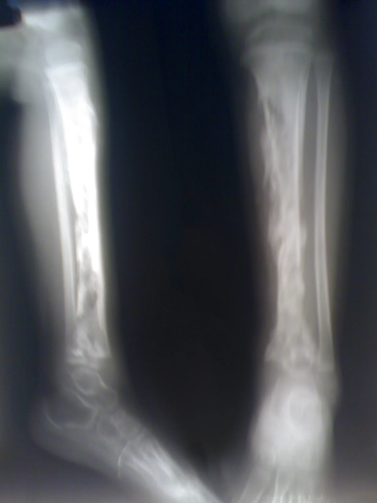
Chronic osteomyelitis develops when acute infection is inadequately treated or the bone's blood supply is compromised: dead bone (a sequestrum) becomes walled off by new bone formation (an involucrum), sheltering bacteria from both antibiotics and the immune system and producing a discharging sinus that can persist or recur for years. A Brodie's abscess is a localised, chronic, sub-acute form, often in the metaphysis of a long bone in a child or young adult, that can mimic a bone tumour radiologically.
Microbiology by context
Staphylococcus aureus is the commonest cause of both osteomyelitis and septic arthritis across almost every age group and setting, and empirical antibiotic choice starts from covering it. Several specific contexts point towards a different organism and are frequently examined.
| Context | Organism to suspect |
|---|---|
| Most patients, any age | Staphylococcus aureus |
| Neonates | Group B Streptococcus, E. coli, S. aureus |
| Children under about 4 years | Kingella kingae, alongside S. aureus |
| Sickle cell disease | Salmonella species - classically over-represented compared with the general population, though S. aureus remains at least as common |
| Sexually active young adults | Neisseria gonorrhoeae - disseminated gonococcal infection, often with a migratory polyarthralgia, tenosynovitis and a sparse pustular rash preceding a single hot joint |
| Puncture wound through a trainer sole | Pseudomonas aeruginosa |
| IV drug use | S. aureus, with a predisposition to unusual sites - the sternoclavicular joint, the pubic symphysis, and vertebral osteomyelitis |
| Prosthetic joint infection | Coagulase-negative staphylococci (e.g. Staphylococcus epidermidis) and S. aureus, often as a biofilm on the implant surface |
Clinical features
Septic arthritis classically presents with a single hot, swollen, exquisitely tender joint with a marked reduction in range of movement - the patient actively resists any passive movement of the joint, which is one of the more reliable bedside signs. Fever and systemic upset are common but can be absent, especially in the very young, the very old, or the immunosuppressed.
Osteomyelitis presents with localised bone pain, tenderness, warmth and swelling over the affected site, with fever in acute disease. In young children, a limp or refusal to weight-bear may be the only clue - acute limp with fever in a child is septic arthritis or osteomyelitis until excluded. Chronic osteomyelitis can present far more insidiously, with a non-healing wound, an intermittently discharging sinus, or low-grade malaise over months.
The Kocher criteria
In children, the Kocher criteria help estimate the probability that an irritable hip is septic arthritis rather than the much commoner and self-limiting transient synovitis.1
| Criterion | Threshold |
|---|---|
| Weight bearing | Unable to weight bear on the affected side |
| Fever | Temperature >38.5°C |
| ESR | >40 mm/hr |
| White cell count | >12 x 10⁹/L |
The probability of septic arthritis rises steeply with the number of criteria met - roughly 3% with none, and over 90% with all four in the original validation cohort - but the score is a probability estimate, not a substitute for aspiration where suspicion remains.
Differential diagnosis
- Transient synovitis - the main paediatric differential, discussed above
- Reactive arthritis - follows a gastrointestinal or genitourinary infection by 1-4 weeks, and the joint itself is sterile
- Gout and pseudogout - can look identical to septic arthritis clinically; joint aspiration with polarised light microscopy is needed to look for crystals, and does not exclude infection on its own since crystal disease and sepsis can coexist
- Haemarthrosis - trauma, anticoagulation, or a bleeding disorder
- Overlying cellulitis without joint involvement - the joint itself moves relatively freely, unlike in septic arthritis
- Perthes' disease and slipped upper femoral epiphysis in children with hip pain - age, X-ray findings and the absence of fever help distinguish these
- Avascular necrosis and bone infarction, particularly in sickle cell disease, which can mimic osteomyelitis clinically and radiologically and often coexists with it
Investigations
Blood tests
- FBC, CRP and ESR - CRP is more useful for tracking response to treatment than for diagnosis alone, since it can be normal early
- Blood cultures - positive in a substantial proportion of both conditions, especially where spread is haematogenous, and should be taken in every case before antibiotics
- Sickle cell screen, HbA1c or other context-specific tests where relevant to the differential
Joint aspiration
Aspirate before starting antibiotics wherever the patient's condition allows it - this is the single most important diagnostic step in suspected septic arthritis. Synovial fluid is sent for Gram stain, culture and sensitivity, white cell count and differential, and polarised light microscopy for crystals.
Imaging
- Plain X-ray - often normal in the first 1-2 weeks of acute osteomyelitis; later signs include periosteal reaction, lytic lesions and, in chronic disease, a visible sequestrum or involucrum. Useful in septic arthritis mainly to exclude a fracture or other bony pathology
- MRI - the most sensitive and specific imaging test for osteomyelitis, and the investigation of choice when it is suspected, since it detects marrow oedema well before plain films change and defines any associated abscess or joint involvement
- Ultrasound - useful for detecting a joint effusion, particularly in the hip, and to guide aspiration
- Bone scan (technetium-99m) - an alternative when MRI is contraindicated or unavailable, useful for identifying multifocal disease
Management
Management combines source control with antibiotics, and the balance between the two differs between the two conditions.
Septic arthritis
Septic arthritis needs surgical washout as well as antibiotics - it is a joint emergency, because untreated infection destroys articular cartilage within days through a combination of bacterial and host inflammatory enzymes. Washout is performed arthroscopically or open, and may need to be repeated. Empirical IV antibiotics are started immediately after aspiration (or alongside it if the patient is septic and cannot wait), typically flucloxacillin, and then rationalised once culture results are available.
Osteomyelitis
Acute osteomyelitis without an abscess or dead bone may respond to antibiotics alone if started early. Where there is an abscess, a sequestrum, or established chronic osteomyelitis, surgical debridement is required alongside antibiotics - antibiotics cannot penetrate dead, avascular bone or clear an established biofilm.
Antibiotic duration and route: the OVIVA trial
Historical practice was 4-6 weeks of IV antibiotics for bone and joint infection, largely on the assumption that oral agents could not achieve adequate bone concentrations. The OVIVA trial (2019) randomised patients with bone or joint infection to IV or oral antibiotics after the first week and found oral therapy non-inferior for treatment failure at 1 year, provided a highly bioavailable oral agent was chosen (for example, a fluoroquinolone, co-trimoxazole, doxycycline or linezolid, depending on the organism).2 This has changed UK practice: many patients now switch to oral antibiotics substantially earlier than 6 weeks of IV therapy once they are clinically stable, a source has been controlled, and an appropriate oral option exists - shortening admissions and avoiding the complications of prolonged IV access.
Prosthetic joint infection
Infection around a prosthetic joint is managed differently, guided by how long after implantation the infection presents and how stable the implant is. Options include DAIR (debridement, antibiotics and implant retention) for early, well-fixed infections, or one- or two-stage revision for established or biofilm-associated infection, always with close orthopaedic and microbiology multidisciplinary input given the long antibiotic courses and complex surgical decisions involved.
Complications
- Joint destruction and secondary osteoarthritis after septic arthritis, especially with any delay to washout
- Growth plate damage and resulting limb length discrepancy or angular deformity in children
- Chronic osteomyelitis, with recurrent flares and discharging sinuses, sometimes for years
- Pathological fracture through weakened bone
- Bacteraemia and sepsis
- Rarely, squamous cell carcinoma arising in a chronically discharging sinus (Marjolin's ulcer) after many years of chronic osteomyelitis
Red flags
Prognosis
Outcomes are good with prompt diagnosis and treatment, particularly in previously healthy patients with acute disease. The strongest predictor of a poor outcome in septic arthritis is delay to joint washout, since cartilage damage begins early and is irreversible. In children, delayed diagnosis risks growth disturbance that only becomes apparent years later. Chronic osteomyelitis, once established, can be difficult to eradicate completely and may require prolonged or repeated treatment over years.
References
- Kocher MS, Zurakowski D, Kasser JR. Differentiating between septic arthritis and transient synovitis of the hip in children: an evidence-based clinical prediction algorithm. Journal of Bone and Joint Surgery (Am). 1999. Available here
- Li HK, Rombach I, Zambellas R et al. Oral versus intravenous antibiotics for bone and joint infection (OVIVA). New England Journal of Medicine. 2019. Available here
- NICE Clinical Knowledge Summaries. Bone and joint infections. Available here
- British Society for Antimicrobial Chemotherapy. Guidance on the diagnosis and management of prosthetic joint infection. Journal of Antimicrobial Chemotherapy. 2021. Available here
- Kremers HM, Nwojo ME, Ransom JE et al. Trends in the epidemiology of osteomyelitis: a population-based study. Journal of Bone and Joint Surgery (Am). 2015. Available here
This article is written for revision and education. It is not clinical guidance and must not be used to make decisions about the care of a patient. Always check current NICE guidance and local protocols.