
Immune Thrombocytopenic Purpura
Key points
- Immune thrombocytopenic purpura (ITP): isolated thrombocytopenia caused by immune-mediated platelet destruction (autoantibodies against platelet surface glycoproteins) and, to a lesser extent, impaired platelet production.
- Primary vs secondary: primary (idiopathic) ITP has no identifiable cause; secondary ITP occurs in the context of another condition - SLE, HIV, hepatitis C, H. pylori infection, lymphoproliferative disease, or certain drugs.
- Two very different patterns: childhood ITP is typically acute, often follows a viral illness, and is usually self-limiting within weeks to months. Adult ITP is typically more chronic, more common in women, and less often resolves spontaneously.
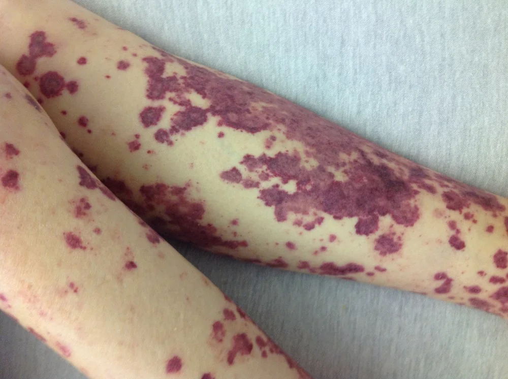
- Clinical features: petechiae, purpura and mucosal bleeding (epistaxis, gum bleeding, menorrhagia) in an otherwise well patient. No lymphadenopathy or organomegaly - their presence should prompt reconsideration of the diagnosis.
- Diagnosis: a diagnosis of exclusion: isolated thrombocytopenia on FBC, a blood film to exclude pseudothrombocytopenia and other causes (schistocytes, blasts), with bone marrow biopsy reserved for atypical presentations, not routine confirmation.
- Management threshold: treatment is guided by bleeding symptoms, not the platelet count alone - many patients, especially children, need only observation. Treat for active bleeding or a platelet count low enough to carry a significant spontaneous bleeding risk.
- First-line treatment: oral corticosteroids; IV immunoglobulin (IVIG) for a faster rise (active bleeding, urgent procedure). Second line: rituximab, splenectomy, or TPO-receptor agonists (eltrombopag, romiplostim) for chronic/refractory disease.
- Feared complication: intracranial haemorrhage - rare, but the reason severe or symptomatic thrombocytopenia is treated promptly rather than simply observed.
Introduction
Immune thrombocytopenic purpura (ITP) is an acquired condition in which antibodies directed against platelet surface glycoproteins (commonly GPIIb/IIIa) cause accelerated platelet destruction by the reticuloendothelial system, chiefly the spleen. Impaired platelet production, from the same antibodies affecting megakaryocytes, also contributes.1
ITP is a diagnosis of exclusion: there is no single confirmatory test, and the diagnosis rests on finding isolated thrombocytopenia with no other identifiable cause, after a careful assessment for secondary causes and mimics.
Classification and aetiology
Primary vs secondary
- Primary (idiopathic) ITP - no identifiable underlying cause; the majority of cases
- Secondary ITP - occurring in association with SLE, HIV, hepatitis C, Helicobacter pylori infection, lymphoproliferative disorders (e.g. CLL), common variable immunodeficiency, or certain drugs (e.g. heparin - though heparin-induced thrombocytopenia is a distinct, prothrombotic entity - quinine, some antibiotics)
Childhood vs adult disease
| Feature | Childhood ITP | Adult ITP |
|---|---|---|
| Onset | Often abrupt, frequently 1-4 weeks after a viral illness | More insidious |
| Sex distribution | Equal | Female predominance |
| Natural history | Usually self-limiting, resolving within weeks to months in the majority | Often becomes chronic (>12 months), less likely to remit spontaneously |
| Typical management | Observation in most cases, even with quite low counts, if no significant bleeding | More often requires treatment |
Clinical features
Patients are typically otherwise well, with bleeding manifestations proportional to how low the platelet count has fallen:
- Petechiae - pinpoint, non-blanching red-purple spots, typically on the lower legs
- Purpura and easy bruising
- Mucosal bleeding - epistaxis, gum bleeding, menorrhagia
- Gastrointestinal or urinary bleeding in more severe thrombocytopenia
- No fever, weight loss, bone pain, lymphadenopathy or organomegaly - their presence points away from ITP
Investigations
- FBC - isolated thrombocytopenia, with other cell lines normal
- Blood film - essential, to exclude pseudothrombocytopenia (EDTA-induced platelet clumping - an artefact, not true thrombocytopenia, confirmed by repeating in a citrate tube), and to look for schistocytes (suggesting TTP/HUS/DIC) or blasts (suggesting leukaemia), both of which would exclude a simple ITP diagnosis
- Clotting screen - typically normal in ITP, unlike DIC
- HIV and hepatitis C serology, and consideration of H. pylori testing and autoimmune screen (ANA) if secondary ITP is suspected
- Bone marrow biopsy is not routinely required. It is reserved for atypical presentations - additional cytopenias, systemic symptoms, an unexpected age group, or poor response to first-line treatment - to exclude leukaemia, aplastic anaemia or myelodysplasia before immunosuppressive treatment is given
- Antiplatelet antibody testing is not routinely used, given limited sensitivity and specificity
Differential diagnosis
- Pseudothrombocytopenia - an EDTA sample artefact; always confirm with a citrate sample or blood film before treating a surprising thrombocytopenia result
- Thrombotic thrombocytopenic purpura / haemolytic uraemic syndrome - thrombocytopenia with schistocytes, haemolysis and (in TTP) neurological signs/renal impairment - a medical emergency requiring urgent specialist management, quite different from ITP
- Disseminated intravascular coagulation - deranged clotting and low fibrinogen, unlike the normal coagulation profile in ITP
- Drug-induced thrombocytopenia (including heparin-induced thrombocytopenia, a prothrombotic condition requiring immediate cessation of heparin)
- Bone marrow failure or infiltration (aplastic anaemia, leukaemia, myelodysplasia) - usually with additional cytopenias or abnormal film findings
- Hypersplenism - splenomegaly present, unlike typical ITP
- Gestational thrombocytopenia in pregnancy - mild, incidental, and not ITP
Management
The decision to treat is based on bleeding symptoms and platelet count together, not the count in isolation - many patients, especially children with an asymptomatic low count, need no treatment at all beyond safety-netting advice and monitoring.2
Children
Observation alone is appropriate for most children with no or minor bleeding, even with quite low platelet counts, given the high likelihood of spontaneous resolution. Treatment is reserved for significant bleeding or very low counts with risk factors, using the same options as in adults.
First-line treatment (significant bleeding or low platelet count)
- Oral corticosteroids (e.g. prednisolone) - first line, producing a response in the majority, though relapse after tapering is common
- IV immunoglobulin (IVIG) - produces a faster rise in platelet count than steroids, useful for active bleeding or before an urgent procedure/surgery, though the effect is often temporary
- Anti-D immunoglobulin - an alternative in Rh-positive, non-splenectomised patients
Second-line/chronic disease
- Rituximab - depletes B cells, reducing autoantibody production
- TPO-receptor agonists (eltrombopag, romiplostim) - stimulate platelet production, an important option particularly for chronic/relapsing disease
- Splenectomy - removes the primary site of both antibody production and platelet destruction; effective but now used less often given the efficacy of medical alternatives, and requires the usual pre-operative vaccination and lifelong consideration of hyposplenism risk
- Treat the underlying cause in secondary ITP where identified (e.g. H. pylori eradication, antiretroviral therapy for HIV)
Emergency management of severe bleeding
IV methylprednisolone, IVIG and platelet transfusion together for life-threatening bleeding (e.g. intracranial haemorrhage) - platelet transfusion alone is usually rapidly consumed by ongoing antibody-mediated destruction and is reserved for genuine emergencies rather than used routinely.
Complications
- Intracranial haemorrhage - rare (well under 1%) but the most feared complication, and the main driver of treatment thresholds in severe thrombocytopenia
- Significant mucosal or gastrointestinal bleeding
- Chronic ITP - persistent disease beyond 12 months, more common in adults
- Treatment-related complications - steroid side effects with prolonged courses, infection risk after splenectomy or rituximab
Red flags
Prognosis
Childhood ITP has an excellent prognosis, with the majority of children achieving spontaneous remission within weeks to a few months, and only a minority progressing to chronic disease.2 Adult ITP is more often chronic, with many patients requiring ongoing or intermittent treatment over years, though the overall risk of serious bleeding remains low with appropriate monitoring and treatment thresholds.
With modern management - including second-line options such as TPO-receptor agonists and rituximab for those who relapse after steroids - the large majority of patients, both children and adults, achieve good long-term control of bleeding risk, and mortality directly attributable to ITP is low.
References
- Neunert C, Terrell DR, Arnold DM et al. American Society of Hematology 2019 guidelines for immune thrombocytopenia. Blood Adv. 2019. Available here
- Provan D, Arnold DM, Bussel JB et al. Updated international consensus report on the investigation and management of primary immune thrombocytopenia. Blood Adv. 2019. Available here
- DrFO.Jr.Tn, CC BY-SA 3.0, via Wikimedia Commons. Available here
- NICE Clinical Knowledge Summaries (CKS). Thrombocytopenia. 2023. Available here
- NHS. Idiopathic thrombocytopenic purpura (ITP). 2023. Available here
This article is written for revision and education. It is not clinical guidance and must not be used to make decisions about the care of a patient. Always check current NICE guidance and local protocols.