
Age-Related Macular Degeneration
Key points
- Definition: degeneration of the macula, causing progressive loss of central vision with preservation of peripheral vision. The commonest cause of blindness in the UK.
- Dry (non-neovascular): around 90% of cases. Gradual central blurring over years, with drusen and retinal pigment epithelial changes, progressing to geographic atrophy.
- Wet (neovascular): around 10% of cases but most of the severe visual loss. Choroidal neovascularisation leaks and bleeds, causing rapid distortion and central loss over weeks.
- The defining symptom: metamorphopsia - straight lines appear wavy or bent. Detected with an Amsler grid, which every at-risk patient should be given.
- Never affects: peripheral vision or navigation. A patient with AMD who cannot find their way around has something else as well.
- Wet AMD treatment: intravitreal anti-VEGF - ranibizumab, aflibercept or bevacizumab - which stabilises vision in around 90% and improves it in about a third.
- Urgency: suspected wet AMD must be seen by a macular service within two weeks, and many units aim for the same week. Delay costs vision permanently.
- Dry AMD: no proven treatment. Stop smoking, control blood pressure, and consider AREDS2 supplements for intermediate or advanced disease in one eye.
Introduction
Age-related macular degeneration is the leading cause of blindness in the UK and in the developed world, accounting for around half of all certifications of severe sight impairment.1 It affects roughly one in ten people over 65 to some degree and one in four over 75.
The macula is the small central area of retina, about 5.5 mm across, containing the fovea at its centre. It has the highest density of cone photoreceptors in the eye and provides all detailed, high-acuity, colour vision - reading, recognising faces, seeing detail. It occupies a tiny fraction of the retinal area but a disproportionate share of the visual cortex.
The consequence is a very characteristic pattern of disability. AMD destroys exactly the vision needed to read, drive and recognise people, while leaving navigational and peripheral vision intact. Patients are therefore severely disabled for close work yet walk into clinic unaided, which is often misinterpreted by families as the problem being less serious than it is. Being able to explain this distinction is a large part of managing the condition well.
Pathophysiology and classification
The disease process begins in the outer retina, at the interface between the photoreceptors, the retinal pigment epithelium (RPE), Bruch's membrane and the choriocapillaris. Photoreceptor outer segments are shed and phagocytosed by the RPE continuously throughout life. With age this process becomes inefficient, and undigested material accumulates as drusen between the RPE and Bruch's membrane, along with lipofuscin within RPE cells.
Drusen thicken and stiffen Bruch's membrane, impairing exchange between the choroidal circulation and the outer retina. The resulting oxidative stress, chronic low-grade inflammation and complement activation - the genetic association with complement factor H is one of the strongest in complex disease - drive RPE dysfunction and photoreceptor loss. From this common starting point, the disease diverges into two forms.
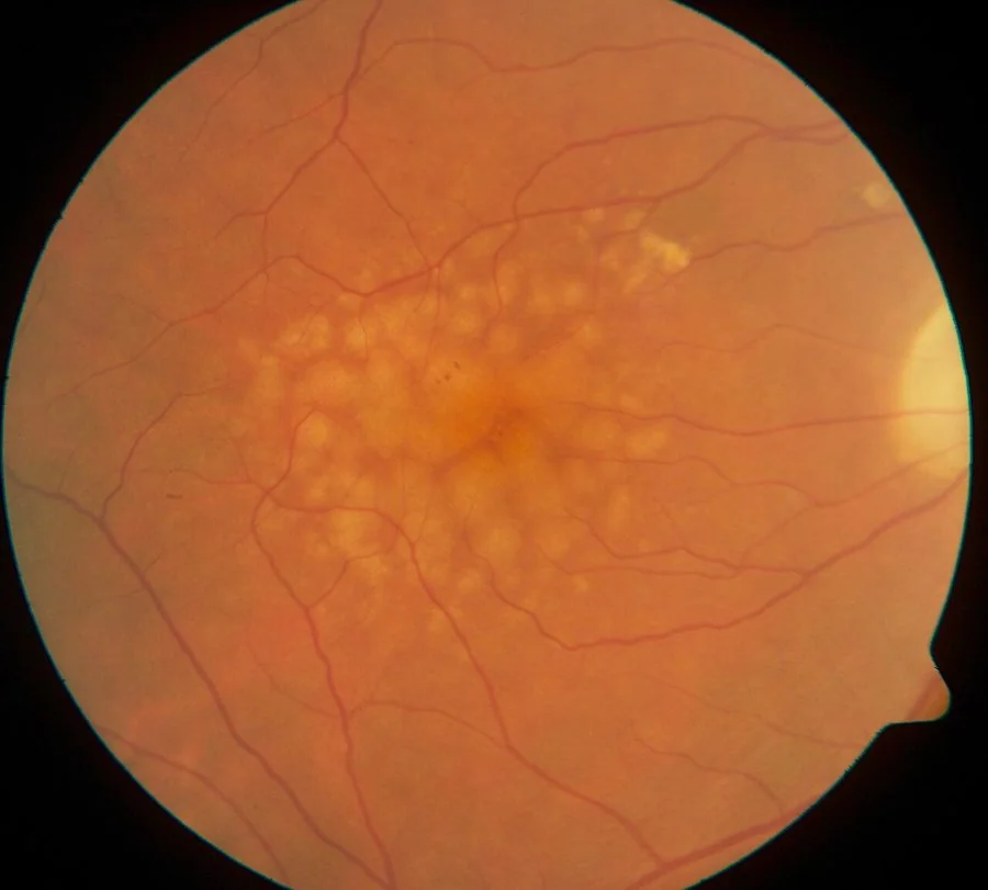
Dry (non-neovascular, atrophic) AMD
Around 90% of cases. Drusen accumulate and RPE cells and their overlying photoreceptors die, gradually over years. Advanced dry AMD is geographic atrophy: sharply demarcated areas of RPE and photoreceptor loss through which the underlying choroidal vessels are visible. Vision declines slowly and there is, until very recently, no treatment that halts it.
- Early AMD - medium-sized drusen (63-125 micrometres) with no pigment abnormality; usually asymptomatic
- Intermediate AMD - large drusen (over 125 micrometres) or pigmentary changes; mild symptoms, and this is the group at meaningful risk of progression
- Advanced dry AMD - geographic atrophy involving the fovea, with a dense central scotoma
Wet (neovascular, exudative) AMD
Around 10% of cases but the cause of most severe and rapid visual loss. Hypoxia and inflammation in the outer retina drive expression of vascular endothelial growth factor (VEGF), which stimulates new vessels to grow from the choriocapillaris through breaks in Bruch's membrane - choroidal neovascularisation. These vessels are fragile and leaky, so they exude fluid and lipid and bleed, lifting and distorting the retina. Untreated, the process ends in a fibrovascular disciform scar with a dense central scotoma, typically within months.
Risk factors
- Increasing age - the dominant risk factor, with prevalence rising steeply after 70
- Smoking - the strongest modifiable risk factor, increasing risk two- to fourfold and bringing forward onset by around a decade. Risk falls after stopping but takes many years to normalise.
- Family history and genetics - variants in complement factor H and ARMS2/HTRA1 confer substantial risk; a first-degree relative roughly triples it
- White European ethnicity - AMD is markedly less common in people of African and Asian ancestry
- Cardiovascular risk factors - hypertension, hypercholesterolaemia and obesity
- Diet - low intake of the carotenoids lutein and zeaxanthin, and of omega-3 fatty acids; high intake of saturated fat
- Female sex, partly reflecting longer life expectancy
- Cumulative ultraviolet and blue light exposure
- Previous cataract surgery - an association reported in some cohorts, though confounded by ascertainment
- Advanced AMD in the fellow eye - the single strongest predictor of what will happen to the second eye
Clinical features
Symptoms
- Gradual or sudden central visual blurring, depending on type
- Metamorphopsia - straight lines appearing wavy, bent or interrupted. Patients describe door frames bowing, lamp posts leaning, or letters running together. This is the hallmark of neovascular disease.
- Central scotoma - a grey, dark or missing patch in the middle of vision, which persists rather than moving with gaze
- Micropsia or macropsia - objects appearing smaller or larger in the affected eye, from displacement of photoreceptors by subretinal fluid
- Difficulty reading, needing brighter light, losing the line, or finding letters missing from words
- Difficulty recognising faces while still seeing the person's outline
- Reduced contrast sensitivity and slow dark adaptation
- Charles Bonnet syndrome - formed visual hallucinations of people, animals or patterns in someone with insight that they are not real
Signs
| Finding | Dry AMD | Wet AMD |
|---|---|---|
| Drusen | Hallmark feature - yellow deposits at the macula; small hard drusen are low risk, large soft confluent drusen high risk | Usually present as a background |
| Pigment | Mottling, clumping and atrophy of the retinal pigment epithelium | May be present |
| Atrophy | Geographic atrophy - sharply edged pale patches with visible choroidal vessels | Not the primary feature |
| Haemorrhage | Absent | Subretinal or intraretinal haemorrhage at the macula |
| Exudate | Absent | Hard exudates, subretinal fluid, retinal or pigment epithelial elevation |
| Scarring | Absent | Grey-green subretinal membrane, progressing to a fibrotic disciform scar |
| Course | Years | Weeks to months |
The Amsler grid
A square grid with a central fixation dot, viewed at normal reading distance with reading glasses on, one eye at a time. The patient fixates the central dot and reports whether any lines are wavy, blurred, broken or missing. It is cheap, takes a minute, and is the standard means of both detecting and monitoring macular disease. Every patient with drusen or dry AMD should be given one, shown how to use it, told to test each eye daily, and told explicitly to seek same-week review if the pattern changes.2
Differential diagnosis
- Diabetic maculopathy - central blurring with microaneurysms, dot and blot haemorrhages and circinate exudates; check the diabetes history and the rest of the fundus
- Cataract - gradual blurring but with dulling of the red reflex, glare and no metamorphopsia
- Central serous chorioretinopathy - a younger patient, typically a man in his 30s or 40s under stress or on corticosteroids, with a serous macular detachment causing micropsia and blurring
- Macular hole - central scotoma with distortion; diagnosed on OCT
- Epiretinal membrane - metamorphopsia from a cellophane-like membrane contracting on the macular surface
- Myopic macular degeneration - choroidal neovascularisation in high myopia, at younger ages
- Retinal vein occlusion with macular oedema - sudden painless blurring with fundal haemorrhages in the affected territory
- Cystoid macular oedema after cataract surgery
- Toxic maculopathy - hydroxychloroquine causing a bullseye maculopathy, which is why patients on long-term treatment are screened
Investigations
- Visual acuity in each eye separately, with a pinhole, and ideally with a logMAR chart for accuracy in tracking change
- Amsler grid for metamorphopsia and scotoma mapping
- Dilated fundus examination with slit lamp biomicroscopy, to identify drusen, pigment change, atrophy, haemorrhage and exudate
- Optical coherence tomography - the key investigation. It is non-invasive, takes seconds, and shows drusen, intraretinal and subretinal fluid, pigment epithelial detachment and atrophy in cross-section. It both makes the diagnosis of wet AMD and tracks the response to each injection.
- OCT angiography - visualises the neovascular membrane without dye injection, increasingly used in place of fluorescein angiography
- Fluorescein angiography - historically the diagnostic standard for choroidal neovascularisation, showing early hyperfluorescence with late leakage; still used where the diagnosis is unclear
- Indocyanine green angiography - for polypoidal choroidal vasculopathy and occult membranes, since it images the choroidal circulation better than fluorescein
Management
Wet AMD: intravitreal anti-VEGF
Anti-VEGF therapy transformed the prognosis of wet AMD. Before it, the natural history was progressive loss to a disciform scar; with it, around 90% of eyes have vision stabilised and roughly a third gain meaningful acuity.4 Agents used in the UK include ranibizumab, aflibercept, faricimab and, off-label but widely used and comparably effective, bevacizumab.
- Given by intravitreal injection under topical anaesthesia in a clean room, taking a few minutes
- A loading phase of three monthly injections, then continued monthly, bimonthly or on a treat-and-extend regimen guided by OCT
- Treatment is usually ongoing for years; stopping often leads to recurrence, and adherence over that timescale is a genuine clinical problem
- Risks of injection - endophthalmitis (around 1 in 2,000-3,000 injections), retinal detachment, raised intraocular pressure, subconjunctival haemorrhage, cataract and, rarely, arterial thromboembolic events
- Photodynamic therapy with verteporfin and thermal laser photocoagulation are now largely historical, retained for specific membrane types such as polypoidal disease
Dry AMD
There is no established treatment that restores vision in dry AMD, and until recently none that slowed it. Management is therefore risk reduction, monitoring for conversion to wet disease, and rehabilitation.
- Smoking cessation - the single most effective intervention, and worth raising at every visit
- Blood pressure and lipid control, and cardiovascular risk management generally
- Diet - a Mediterranean-style diet with oily fish and dark green leafy vegetables rich in lutein and zeaxanthin
- AREDS2 supplementation - vitamins C and E, zinc, copper, lutein and zeaxanthin reduced progression to advanced AMD by around 25% over five years in those with intermediate AMD in both eyes or advanced AMD in one eye. It has no proven benefit in early disease or in people with healthy maculae. Beta-carotene was removed from the original formulation because it increased lung cancer risk in smokers.5
- Amsler grid monitoring with clear instructions on when to seek help
- Complement inhibitors - intravitreal pegcetacoplan and avacincaptad pegol slow the growth of geographic atrophy in trials, but at the time of writing are not approved for NHS use and their functional benefit remains debated
Supportive and rehabilitative care
- Low vision assessment - magnifiers, telescopes, electronic video magnifiers and task lighting
- Certification of visual impairment - offer registration as sight impaired or severely sight impaired, which unlocks Blind Person's Allowance, council tax reduction and social services support
- Practical adaptation - large-print and audiobooks, talking watches and scales, high-contrast markings on steps and switches, and eccentric viewing training to use a preferred retinal locus outside the scotoma
- Driving - patients must meet the DVLA visual standards and must notify the DVLA if they no longer do
- Psychological support - depression is common and under-recognised, and loss of reading and driving is often experienced as a loss of independence
- Charitable support - the Macular Society and RNIB provide counselling, peer support groups and practical advice
Prognosis
Dry AMD progresses slowly. Most people with early or intermediate disease retain useful reading vision for many years, and only a minority progress to foveal geographic atrophy. The risk of progression rises with the number and size of drusen, the presence of pigmentary change, and advanced disease in the fellow eye. Around 10-15% of eyes with dry AMD convert to wet AMD, which is why monitoring matters.
Untreated wet AMD has a poor prognosis, with most eyes losing three or more lines of acuity within a year and progressing to a disciform scar. With anti-VEGF started promptly, vision is stabilised in the great majority. The strongest predictor of the final result is the acuity at which treatment starts, which is the entire rationale for the two-week referral standard: vision already lost to subretinal fibrosis does not come back, whereas fluid resolves.
Whatever the type, it is worth stating explicitly to patients that AMD does not cause total blindness. Peripheral vision is retained, so people continue to move around independently, recognise shapes and manage at home. The disability is severe and specific - reading, faces, driving - and framing it accurately, rather than allowing a patient to leave believing they are going to lose all sight, is one of the more valuable things done in that consultation.
References
- Royal College of Ophthalmologists. Age-Related Macular Degeneration: guidelines for management. Available here
- NICE Clinical Knowledge Summaries. Macular degeneration - age-related. Available here
- NICE NG82. Age-related macular degeneration. 2018. Available here
- Rosenfeld PJ, Brown DM, Heier JS et al. Ranibizumab for neovascular age-related macular degeneration (MARINA). New England Journal of Medicine. 2006. Available here
- Age-Related Eye Disease Study 2 (AREDS2) Research Group. Lutein plus zeaxanthin and omega-3 fatty acids for age-related macular degeneration. JAMA. 2013. Available here
This article is written for revision and education. It is not clinical guidance and must not be used to make decisions about the care of a patient. Always check current NICE guidance and local protocols.