
Chronic Open Angle Glaucoma
Key points
- Definition: a chronic optic neuropathy with characteristic disc cupping and visual field loss, in the presence of an anatomically open drainage angle.
- Why it is dangerous: it is completely asymptomatic until late. Peripheral field is lost first and the fellow eye compensates, so patients present with irreversible damage.
- Pressure is a risk factor, not the definition: up to 40% of patients have intraocular pressure in the normal range (normal tension glaucoma), and most people with raised pressure never develop glaucoma.
- The disc: increased cup-to-disc ratio (>0.7), asymmetry between the eyes, thinning of the neuroretinal rim, notching, bayoneting of vessels and disc haemorrhages.
- The field: arcuate (Bjerrum) scotoma and a nasal step early, progressing to tunnel vision with central acuity preserved until the very end.
- First-line treatment: selective laser trabeculoplasty (SLT), which NICE now recommends ahead of drops for most newly diagnosed patients.
- Medical treatment: prostaglandin analogue such as latanoprost once nightly, then a beta-blocker, alpha agonist or carbonic anhydrase inhibitor added.
- Driving and registration: significant bilateral field loss must be notified to the DVLA, and eligible patients should be offered certification of visual impairment.
Introduction
Chronic open angle glaucoma - also called primary open angle glaucoma - is a progressive optic neuropathy in which retinal ganglion cell axons are lost, producing characteristic cupping of the optic disc and a corresponding pattern of visual field loss. It affects around 2% of people over 40 in the UK and is one of the leading causes of irreversible blindness worldwide.1
Two features define its clinical character. First, it is painless and asymptomatic until it is advanced: peripheral field is lost gradually, the fellow eye fills in the gap, and central acuity is preserved almost to the end, so patients notice nothing. Around half of people with glaucoma in the UK are undiagnosed, and the majority of those who are diagnosed are picked up incidentally at a routine sight test rather than by presenting with a symptom.2
Second, the damage is irreversible. Retinal ganglion cells do not regenerate, so every treatment in glaucoma is aimed at slowing further loss rather than recovering what has gone. That combination - silent, progressive, irreversible - is what makes case-finding through community optometry the central plank of glaucoma care in the UK, and why the free NHS sight test for people over 40 with a family history exists.
Pathophysiology
Aqueous humour is produced by the ciliary epithelium, flows through the pupil into the anterior chamber, and drains chiefly through the trabecular meshwork into Schlemm's canal, with a smaller proportion leaving by the uveoscleral route. In open angle glaucoma the drainage angle is anatomically open on gonioscopy, but resistance within the trabecular meshwork itself is increased - by extracellular matrix accumulation, loss of trabecular cells and reduced pore size - so pressure rises.
The damage occurs at the lamina cribrosa, the sieve-like plate of collagen through which around a million ganglion cell axons leave the eye. Raised pressure deforms and compresses this structure, obstructing axoplasmic transport and depriving ganglion cells of neurotrophic factors supplied from the brain. Axons die, the neuroretinal rim thins, and the physiological cup enlarges to occupy the space.
The pattern of field loss follows the anatomy of the retinal nerve fibre layer. Axons from the superior and inferior retina arch around the macula in bundles, entering the disc at its superior and inferior poles, which are the regions most vulnerable to laminar deformation. Damage to an arcuate bundle produces the characteristic arcuate scotoma sweeping from the blind spot around the fixation point. Fibres from the nasal retina run straight to the disc and are spared until late, which is why central vision survives so long.
Risk factors
- Raised intraocular pressure - the only modifiable risk factor, and the strength of the association rises steeply above 21 mmHg
- Increasing age - prevalence rises from around 1% at 40 to nearly 10% over 75
- Family history - a first-degree relative increases risk roughly four- to ninefold. Siblings of an affected patient should have annual screening from age 40, funded by the NHS.
- Black African or African-Caribbean ancestry - four to five times the risk, with earlier onset and more aggressive disease
- Myopia - short-sightedness, in contrast to angle closure which is associated with hypermetropia
- Central corneal thickness - a thin cornea both underestimates true pressure on applanation and is an independent risk factor
- Diabetes mellitus and systemic hypertension
- Nocturnal systemic hypotension, including from over-treated hypertension, which reduces optic nerve head perfusion
- Corticosteroid use - topical, inhaled, nasal or systemic, in the roughly one third of the population who are steroid responders
- Pseudoexfoliation and pigment dispersion syndromes, which cause secondary open angle glaucoma
Clinical features
The defining clinical feature is the absence of clinical features. There is no pain, no redness, no discharge and no early change in acuity. When symptoms do appear they are late:
- Gradual peripheral field loss, described as bumping into door frames, missing steps, or difficulty in traffic
- Tunnel vision in advanced disease, with central acuity often still 6/6
- Difficulty with dark adaptation and with glare
- Loss of central vision only at the very end, when the papillomacular bundle finally fails
- Incidental detection at a routine optometry sight test, which is how the majority present
The optic disc
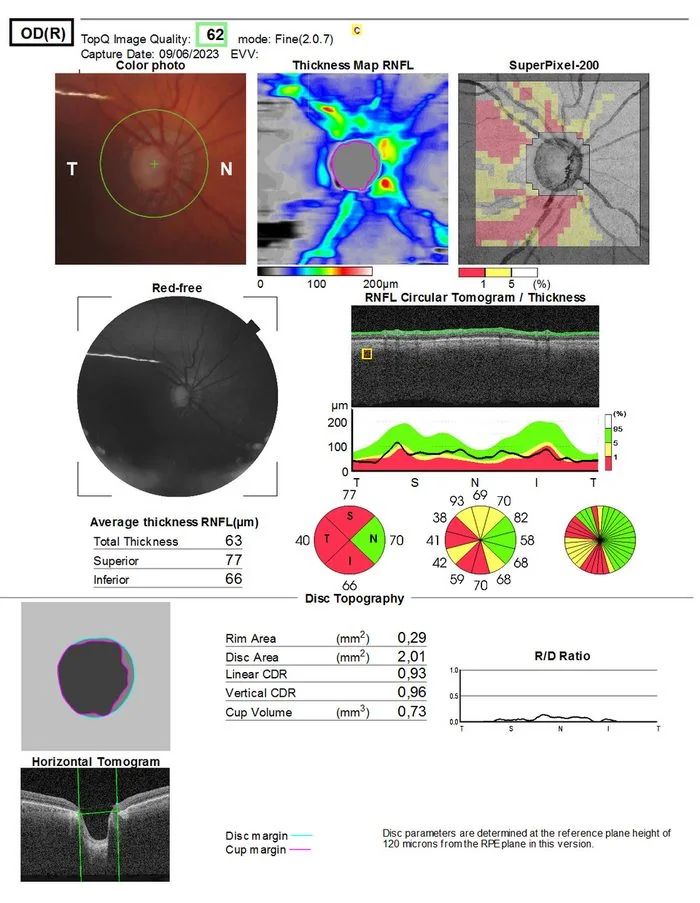
Examining the disc well is a core clinical skill and an examinable one. The cup is the pale central depression; the neuroretinal rim is the orange tissue between the cup and the disc margin, and it contains the axons. What matters is the rim, not the cup.
- Increased cup-to-disc ratio - a ratio above 0.7 is suspicious, but a large disc can have a large cup and be normal, which is why rim area matters more than ratio
- Asymmetry between the eyes - a difference in cup-to-disc ratio of more than 0.2 between eyes is significant, since discs are usually symmetrical
- Violation of the ISNT rule - in a normal disc the rim is thickest Inferiorly, then Superiorly, then Nasally, then Temporally. Loss of this pattern suggests glaucoma.
- Notching - a localised defect in the rim, most often at the inferotemporal or superotemporal pole
- Bayoneting of vessels - retinal vessels appear to bend sharply as they dip into a deep, steep-walled cup
- Baring of circumlinear vessels - a vessel that formerly hugged the cup margin left standing away from it
- Disc (Drance) haemorrhage - a splinter haemorrhage crossing the disc margin, indicating active, progressing disease and predicting further field loss
- Peripapillary atrophy and nerve fibre layer defects visible as dark wedge-shaped striations
The visual field
- Paracentral scotomas and a nasal step - the earliest detectable changes, in the Bjerrum area 10-20° from fixation
- Arcuate (Bjerrum) scotoma - an arc extending from the blind spot around fixation, respecting the horizontal midline because superior and inferior fibre bundles do not cross it
- Ring scotoma - when superior and inferior arcuate defects join
- Tunnel vision - progressive constriction leaving a small central island and a temporal crescent
- Complete loss - the temporal island being typically the last to go
Investigations
NICE specifies a defined set of tests for diagnosis and monitoring, and the same set is repeated at follow-up to detect progression.3
| Investigation | What it measures | Notes |
|---|---|---|
| Goldmann applanation tonometry | Intraocular pressure | The reference standard. Non-contact tonometry is used for screening but is less accurate. |
| Central corneal thickness (pachymetry) | Corneal thickness | A thin cornea causes underestimation of true pressure and is an independent risk factor; a thick cornea causes overestimation |
| Gonioscopy | The drainage angle | Confirms the angle is open, distinguishing this from angle closure and identifying secondary causes such as pigment or neovascularisation |
| Standard automated perimetry | Visual field | Usually Humphrey 24-2. Provides the functional measure of damage and the primary means of tracking progression. |
| Optic nerve head assessment | Structure | Stereoscopic slit lamp examination with a fundus lens, plus disc photography for comparison over time |
| Optical coherence tomography | Retinal nerve fibre layer and ganglion cell thickness | Detects structural loss before field loss becomes apparent, and quantifies progression objectively |
Management
The aim of all treatment is to lower intraocular pressure sufficiently to halt or slow progression, expressed as an individualised target pressure based on the baseline pressure, the severity of existing damage, life expectancy and rate of progression. A typical target is a 20-30% reduction from baseline.
Selective laser trabeculoplasty
NICE now recommends SLT as first-line treatment for most people with newly diagnosed open angle glaucoma or ocular hypertension, before drops.3 This followed the LiGHT trial, which found that SLT gave at least equivalent pressure control and quality of life compared with drops, left more patients drop-free at three years, reduced the need for glaucoma surgery, and was more cost-effective.4
SLT uses a frequency-doubled Nd:YAG laser to target pigmented trabecular meshwork cells selectively, triggering a biological remodelling response that improves outflow without causing thermal scarring. It is an outpatient procedure taking a few minutes, and it can be repeated. Its effect wanes over three to five years in many patients, who then need repeat treatment or drops.
Topical medical therapy
Where SLT is declined, is unsuitable, or has failed to reach target, treatment starts with a single agent and escalates by adding rather than substituting, provided the first drug produced some benefit. A prostaglandin analogue is first line in almost all cases because it is the most effective class, is given once daily, and has no systemic contraindications.6
| Class | Example | Mechanism | Key adverse effects |
|---|---|---|---|
| Prostaglandin analogue | Latanoprost, once at night | Increases uveoscleral outflow; the most effective single class, lowering pressure by 25-33% | Iris and periocular skin hyperpigmentation, eyelash lengthening, deepening of the upper lid sulcus, conjunctival hyperaemia, and reactivation of herpetic keratitis |
| Beta-blocker | Timolol, twice daily | Reduces aqueous production | Bronchospasm, bradycardia, heart block, fatigue and masking of hypoglycaemia - contraindicated in asthma |
| Alpha-2 agonist | Brimonidine, twice daily | Reduces production and increases uveoscleral outflow | Allergic conjunctivitis, dry mouth, drowsiness. Contraindicated in infants because it causes apnoea. |
| Carbonic anhydrase inhibitor | Dorzolamide, three times daily; oral acetazolamide | Reduces aqueous production | Bitter taste, stinging; oral form causes paraesthesiae, malaise, renal stones and metabolic acidosis |
| Miotic | Pilocarpine, four times daily | Contracts the ciliary muscle, opening the meshwork | Brow ache, induced myopia, small pupil impairing vision in dim light; now rarely used |
| Rho kinase inhibitor | Netarsudil | Increases trabecular outflow | Conjunctival hyperaemia, corneal verticillata; not yet widely used in the UK |
Surgical options
- Trabeculectomy - the standard operation, creating a guarded fistula from the anterior chamber to a subconjunctival bleb. Usually augmented with mitomycin C to prevent scarring. Complications include hypotony, bleb leak, cataract and, rarely, late bleb-related endophthalmitis, which is why these patients are told to report a red eye urgently.
- Glaucoma drainage devices - Baerveldt or Ahmed tube shunts, for refractory disease or where conjunctiva is scarred
- Minimally invasive glaucoma surgery (MIGS) - iStent, Hydrus or XEN, often combined with cataract surgery, with a better safety profile but more modest pressure reduction
- Cyclodiode laser - ablating the ciliary body to reduce aqueous production, in advanced or refractory disease
- Cataract surgery alone - lowers pressure modestly and is often the first surgical step in a patient who also has cataract
Wider management
- Adherence support - roughly half of patients are non-adherent with drops within a year, so ask about it directly and without judgement, simplify regimens, and consider preservative-free preparations for ocular surface intolerance
- Screen first-degree relatives - siblings and children over 40 are entitled to free NHS sight tests
- DVLA notification - required where there is field loss in both eyes; the patient must meet the visual field standard, assessed by an Esterman binocular field test5
- Certification of visual impairment - offer registration as sight impaired or severely sight impaired where the criteria are met, which unlocks practical and financial support
- Low vision services - magnifiers, lighting advice, home hazard assessment and falls prevention
Complications and prognosis
With early detection and adequate pressure control most patients will not go blind in their lifetime. Perhaps 5-10% of treated patients still progress to significant bilateral visual impairment, and the risk is concentrated in those who present late, those with normal tension glaucoma that continues to progress at target pressures, and those who are non-adherent with treatment.
Prognostic factors include the severity of field loss at diagnosis, the rate of progression on serial fields, the level of pressure achieved, disc haemorrhages, and age. A patient diagnosed at 75 with early disease is unlikely to become visually disabled; a patient diagnosed at 45 with advanced disease requires aggressive treatment because they have decades of potential progression ahead.
The functional consequences matter as much as the field chart. Glaucomatous field loss roughly doubles the risk of falls and of motor vehicle collisions, impairs reading speed even with preserved acuity, and is associated with depression and loss of independence. Glaucoma care that stops at the pressure reading has done only half the job; asking about mobility, driving, reading and mood is the other half.
References
- Tham YC, Li X, Wong TY et al. Global prevalence of glaucoma and projections of glaucoma burden through 2040. Ophthalmology. 2014. Available here
- NICE Clinical Knowledge Summaries. Glaucoma. Available here
- NICE NG81. Glaucoma: diagnosis and management. 2017, updated 2022. Available here
- Gazzard G, Konstantakopoulou E, Garway-Heath D et al. Selective laser trabeculoplasty versus eye drops for first-line treatment of ocular hypertension and glaucoma (LiGHT): a multicentre randomised controlled trial. The Lancet. 2019. Available here
- DVLA. Assessing fitness to drive: a guide for medical professionals. Available here
- BNF. Treatment summary: glaucoma and ocular hypertension. Available here
This article is written for revision and education. It is not clinical guidance and must not be used to make decisions about the care of a patient. Always check current NICE guidance and local protocols.