
Fracture X-ray Interpretation
Key points
- The rule of twos: two views, two joints, two sides and two occasions. Most missed fractures are missed because one of these was skipped.
- One view is one view too few: orthogonal views are mandatory - a posterior shoulder dislocation and a mild slipped epiphysis are both invisible on a single projection.
- ABCS: Adequacy and Alignment, Bones, Cartilage and joints, Soft tissues - a repeatable search pattern that stops you stopping at the first abnormality.
- Describe the distal fragment: displacement, angulation and rotation are always stated in terms of the distal fragment relative to the proximal one.
- Soft tissue signs: an elevated posterior fat pad at the elbow, a lipohaemarthrosis at the knee, or prevertebral swelling in the neck all indicate a fracture you may not be able to see.
- Salter-Harris: SALTR - Slipped, Above, Lower, Through, cRush. Type II is commonest; types III to V carry the greatest risk of growth arrest.
- A normal film does not exclude a fracture: up to 30% of scaphoid fractures and a proportion of hip fractures are invisible initially - immobilise and re-image, or arrange MRI.
- Look for the second injury: an isolated fracture of one forearm bone, or of one pelvic ring component, is rarely isolated.
Introduction
Interpreting a musculoskeletal radiograph is one of the few practical skills you will use on your first day as a doctor and every day thereafter. It is also one where errors are common and consequential: missed fractures are among the most frequent causes of diagnostic error in emergency departments, and the great majority are missed not because the fracture was subtle but because the film was not examined systematically.1
Two habits prevent most of these errors. The first is having a fixed search pattern that you apply to every film, so that finding one abnormality does not stop you looking for the second. The second is describing what you see out loud or in writing in a standard format, because the discipline of describing forces you to look at things you would otherwise skip.
Before you look at the bones
- Confirm the patient - name, date of birth, and that this is the film you think it is. Comparing the wrong patient's films is a real and recurring error.
- Check the side marker and the date and time the film was taken
- Identify the projection - AP or PA, lateral, oblique, or a specialist view
- Assess adequacy - is the whole region included, is the exposure reasonable, is the patient rotated? An inadequate film should be repeated, not squinted at.5
A systematic search: ABCS
A - Adequacy and Alignment
Having confirmed adequacy, check that the normal anatomical relationships are preserved. Alignment abnormalities are often more conspicuous than the fracture itself, and several well-known lines exist precisely because they make subtle displacement obvious.
| Line | Where | Normal finding |
|---|---|---|
| Anterior humeral line | Lateral elbow, paediatric | A line down the anterior humeral cortex should pass through the middle third of the capitellum. If it passes anteriorly, suspect a supracondylar fracture with posterior displacement. |
| Radiocapitellar line | Any elbow view | A line along the radial neck should always pass through the capitellum in every projection. If it does not, the radial head is dislocated - the key to spotting a Monteggia injury. |
| Shenton's line | AP pelvis | A smooth continuous curve from the inferior border of the superior pubic ramus along the inferomedial femoral neck. Disruption suggests a femoral neck fracture or hip dislocation. |
| Klein's line | AP pelvis, adolescent | Drawn along the superior femoral neck, it should intersect part of the epiphysis. If it does not, suspect a slipped upper femoral epiphysis. |
| The three cervical lines | Lateral cervical spine | Anterior vertebral, posterior vertebral and spinolaminar lines should each form a smooth curve without steps |
| The three pelvic rings | AP pelvis | The main pelvic ring and the two obturator foramina. A ring rarely breaks in one place - find the second break. |
B - Bones
Trace the entire cortex of every bone on the film, following it round like a road, looking for a step, a break, a buckle or a change in direction. Then look at the trabecular pattern within the bone for a band of sclerosis (impaction) or lucency (a fracture line). Assess overall bone density and texture - and look for any focal lesion, which raises the possibility of a pathological fracture.
C - Cartilage and joints
Assess joint spaces for symmetry and width, check that articular surfaces are congruent and parallel, and confirm nothing is subluxed or dislocated. In children, examine the growth plates for widening or asymmetry, and know which ossification centres should be present for the age.
S - Soft tissues
This step is skipped most often and yields the most. Look for swelling, loss of normal fat planes, foreign bodies, subcutaneous gas, and joint effusions.
Describing a fracture
This is what you will be asked to do in an OSCE, in a written paper, and on the phone to the orthopaedic registrar. Use the same structure every time.
| Element | What to say |
|---|---|
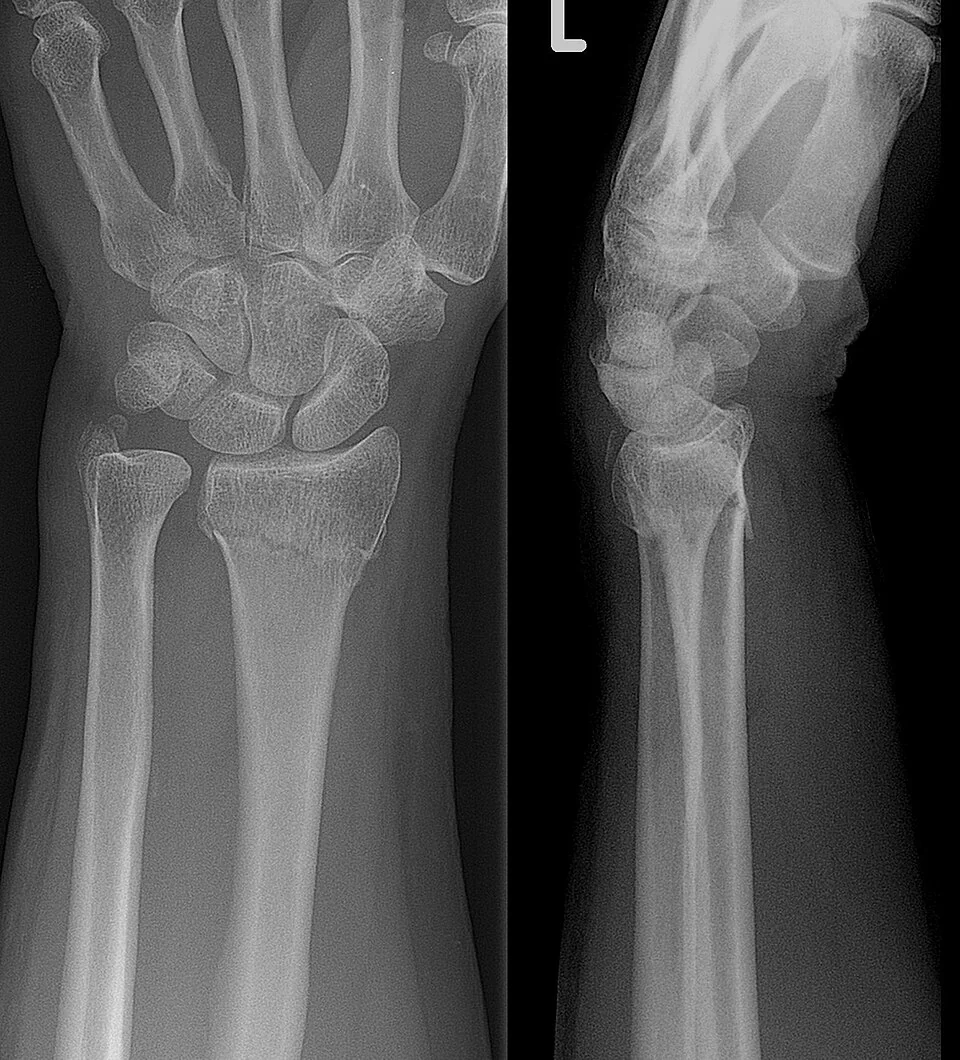
| 1. The film | "This is an AP and lateral radiograph of the left wrist of an adult, taken today" |
| 2. Bone and side | Which bone, which side |
| 3. Location | Proximal, middle or distal third; or epiphysis, metaphysis or diaphysis. State whether it is intra-articular or extra-articular, which is the single most important prognostic feature. |
| 4. Pattern | Transverse, oblique, spiral, comminuted (more than two fragments), segmental, avulsion, impacted; or in children greenstick, buckle (torus) or plastic bowing |
| 5. Displacement | Always describe the distal fragment relative to the proximal. Translation as a percentage of bone width and in which direction; angulation stating the direction the distal fragment points (or the direction the apex points, if you say so explicitly); rotation; and shortening. |
| 6. Joint involvement | Does the fracture enter a joint, is the articular surface stepped, and is the joint dislocated or subluxed? |
| 7. Growth plate | In children, classify by Salter-Harris |
| 8. Other features | Soft tissue swelling, air, foreign bodies, an underlying bone lesion, other fractures, and any metalwork |
| 9. Open or closed | This is a clinical, not a radiological, judgement - it depends on whether the skin is breached, which you determine at the bedside |
Salter-Harris classification
Applies to fractures involving the physis in a skeletally immature patient.4 Remember it as SALTR, and note that the risk of growth disturbance rises with the grade.
| Type | Mnemonic | Anatomy | Growth arrest risk |
|---|---|---|---|
| I | Slipped | Through the physis only. Radiographs may be normal apart from physeal widening - diagnosed clinically by tenderness over the growth plate. A slipped upper femoral epiphysis is essentially a type I. | Low |
| II | Above | Through the physis and up into the metaphysis, taking a triangular metaphyseal fragment (the Thurston-Holland fragment). The commonest type, around 75%. | Low |
| III | Lower | Through the physis and down into the epiphysis - therefore intra-articular | Higher; needs accurate reduction |
| IV | Through | Through metaphysis, physis and epiphysis - intra-articular and crossing the plate | High; usually needs internal fixation |
| V | cRush | Crush injury of the physis. Often diagnosed retrospectively when growth arrest occurs, as the initial film may look normal. | Highest |
Patterns worth recognising
| Fracture | Mechanism and features |
|---|---|
| Colles' | Extra-articular distal radius fracture with dorsal displacement and angulation, from a fall on an outstretched hand. Produces the "dinner fork" deformity. Often with an ulnar styloid fracture. |
| Smith's | The reverse - distal radius with volar displacement, from a fall onto the flexed wrist. Unstable, and usually needs fixation. |
| Barton's | Intra-articular distal radius fracture with subluxation of the carpus |
| Scaphoid | Fall on outstretched hand, tenderness in the anatomical snuffbox, over the scaphoid tubercle, and on axial compression of the thumb. Needs dedicated scaphoid views. |
| Monteggia | Fracture of the proximal ulna with dislocation of the radial head. Check the radiocapitellar line. |
| Galeazzi | Fracture of the distal radius with disruption of the distal radioulnar joint |
| Supracondylar humerus | The commonest paediatric elbow fracture. Check the anterior humeral line, and assess the brachial artery and the median (particularly anterior interosseous) nerve before and after any manipulation. |
| Neck of femur | Classified as intracapsular or extracapsular, which determines whether the blood supply to the head is compromised and therefore whether the hip is fixed or replaced. Look at Shenton's line and the trabecular pattern. |
| Weber ankle classification | By the level of the fibular fracture relative to the syndesmosis: A below (stable), B at the level (variable), C above (unstable, syndesmosis disrupted) |
| Maisonneuve | A proximal fibular fracture with a medial ankle injury - which is why an apparently isolated medial malleolar injury requires the whole fibula to be imaged |
| Lisfranc | Midfoot injury with widening between the first and second metatarsal bases and loss of alignment of the second metatarsal with the middle cuneiform. Frequently missed; consider weight-bearing views or CT. |
Radiographs in children
- Different fracture patterns - the more plastic paediatric bone produces greenstick fractures (one cortex broken, the other bent), buckle or torus fractures (a cortical bulge without a break, usually at the distal radial metaphysis) and plastic bowing with no visible cortical break at all
- Growth plates mimic fractures and vice versa; compare with the opposite side and know the expected appearance for the age
- Elbow ossification centres appear in a fixed order - CRITOE: Capitellum at about 1 year, Radial head at 3, Internal (medial) epicondyle at 5, Trochlea at 7, Olecranon at 9 and External (lateral) epicondyle at 11. If the trochlea is visible but the medial epicondyle is not, the medial epicondyle has been avulsed and displaced - possibly into the joint.
- Pulled elbow (radial head subluxation) in a toddler produces a normal radiograph - the diagnosis is clinical, from the mechanism of a longitudinal pull on a pronated extended arm
- Toddler's fracture - an undisplaced spiral fracture of the tibia in a newly walking child, often invisible initially
Commonly missed fractures
| Missed injury | The clue |
|---|---|
| Scaphoid fracture | Up to 30% are invisible initially. Snuffbox tenderness after a fall on an outstretched hand means immobilise and re-image at 10 to 14 days, or arrange MRI. |
| Radial head fracture | Often visible only as an elevated posterior fat pad. Treat an effusion after elbow trauma as a fracture until proven otherwise. |
| Posterior shoulder dislocation | Invisible on a single AP film. Look for the light bulb sign (internal rotation of the humeral head) and always obtain an axillary or scapular Y view. Classically follows a seizure or electric shock. |
| Occult hip fracture | A patient who cannot weight bear after a fall with a normal radiograph needs MRI - NICE recommends MRI, with CT if MRI is unavailable or cannot be done within 24 hours.3 |
| Impacted femoral neck fracture | No cortical break; look for a band of sclerosis across the neck, disruption of the trabecular pattern, and a broken Shenton's line |
| Cervical spine injury | Inadequate films - the whole of C7 and the C7/T1 junction must be visualised. Use the three lines and the prevertebral soft tissues. |
| Second injury in the forearm or pelvis | A ring or a paired-bone structure rarely breaks in only one place - look for the dislocation or the second fracture |
| Lisfranc and other midfoot injuries | Subtle malalignment of metatarsal bases; consider weight-bearing views or CT if there is midfoot bruising and tenderness |
| Pathological fracture | A trivial mechanism, an unusual site, an underlying lucent or sclerotic lesion, cortical destruction or periosteal reaction. Ask why this bone broke. |
Clinical priorities alongside the film
The radiograph is only part of the assessment, and several things take priority over interpreting it.
- Assess and document neurovascular status before and after any manipulation - pulses, capillary refill, sensation and motor function in the relevant nerve distributions. This is both good practice and an important medicolegal record.
- Open fractures - the wound is photographed, covered with a saline-soaked dressing, the limb is splinted, and intravenous antibiotics and tetanus prophylaxis are given, with urgent orthopaedic and plastic surgical involvement2,7
- Compartment syndrome is diagnosed clinically, not radiologically. Pain out of proportion and pain on passive stretch of the compartment are the key findings; the presence of a pulse does not exclude it.
- Reduce and splint dislocations urgently, particularly where there is skin tenting or neurovascular compromise
- Analgesia early, including regional blocks such as a fascia iliaca block for a hip fracture
- Ask why the bone broke - a low-energy fracture in an older adult should trigger a fracture liaison service referral and osteoporosis assessment, and one in an unusual site should raise the possibility of malignancy
Putting it together
A workable routine takes about a minute. Confirm the patient and the views. Run through ABCS. Apply the rule of twos and ask whether you have the views and the coverage you need. Then describe what you have found in the standard format, and state explicitly what you would do next.
The most useful habit of all is to say what you cannot exclude. "There is no fracture visible on these views, but the patient is tender in the anatomical snuffbox, so I have immobilised the wrist in a splint and arranged repeat imaging in 10 days" is a far better answer than "the X-ray is normal" - and it is the one that protects the patient.
Finally, radiology reports arrive after the patient has often been discharged, and a substantial proportion of missed fractures are actually detected by the reporting radiologist and then lost in the system. Knowing how your department handles discrepant reports, and checking the reports on patients you have sent home, is part of the skill rather than an administrative extra.
References
- NICE NG38. Fractures (non-complex): assessment and management. 2016. Available here
- NICE NG37. Fractures (complex): assessment and management. 2016. Available here
- NICE CG124. Hip fracture: management. 2011, updated 2023. Available here
- Salter RB, Harris WR. Injuries involving the epiphyseal plate. Journal of Bone and Joint Surgery. 1963. Available here
- Royal College of Radiologists. iRefer: making the best use of clinical radiology. Available here
- Royal College of Paediatrics and Child Health. Child Protection Evidence: fractures. Available here
- British Orthopaedic Association Standards for Trauma (BOAST). Open fracture management. Available here
This article is written for revision and education. It is not clinical guidance and must not be used to make decisions about the care of a patient. Always check current NICE guidance and local protocols.