
Otitis Externa: Diagnosis and Management
Key points
- Otitis externa: inflammation of the external auditory canal, usually infective, causing otalgia, itch and discharge.
- Aetiology: most commonly bacterial (Pseudomonas aeruginosa, Staphylococcus aureus); fungal (otomycosis) and eczematous causes also occur.
- Key discriminator: pain on moving the tragus or pinna, which is not a feature of otitis media.
- Risk factors: swimming, humid climates, cotton bud use, hearing aids or earplugs, eczema, and diabetes.
- Management: aural toilet plus a topical antibiotic/steroid preparation; oral antibiotics are not routinely needed.
- Malignant otitis externa: a rare, life-threatening skull base osteomyelitis seen almost exclusively in diabetic or immunocompromised patients - a red flag not to miss.
- Investigations: usually a clinical diagnosis; swab only if resistant, recurrent, or atypical.
- Prognosis: excellent with topical treatment in uncomplicated cases, resolving within 1-2 weeks.
Introduction
Otitis externa is inflammation of the skin lining the external auditory canal, ranging from a localised furuncle to diffuse canal involvement affecting the whole ear.1 It is extremely common, accounting for a large share of ENT presentations in primary care, and is often precipitated by swimming or minor trauma to the canal skin.
The condition is usually straightforward to treat with topical therapy, but it is worth knowing well because it is frequently confused with otitis media, and because a rare but life-threatening variant - malignant (necrotising) otitis externa - can mimic a simple infection in its early stages.2
Aetiology and pathophysiology
The external auditory canal is a warm, enclosed space lined by skin that produces cerumen (ear wax), which is mildly acidic and has some antimicrobial properties. Anything that disrupts this environment - excess moisture, trauma to the skin, or removal of protective wax - allows bacterial or fungal overgrowth and inflammation.1
Bacterial
Pseudomonas aeruginosa and Staphylococcus aureus are the most common organisms, together accounting for the majority of cases. Pseudomonas thrives in the moist environment created by swimming, which is why the condition is colloquially known as "swimmer's ear".1
Fungal (otomycosis)
Aspergillus and Candida species cause otomycosis, which should be suspected when infection is resistant to standard antibacterial drops, occurs after prolonged or repeated antibiotic ear drop use, or when white or black fungal debris is seen on otoscopy.3
Eczematous and other causes
Seborrhoeic dermatitis, atopic eczema and allergic contact dermatitis (from ear drops, jewellery or hearing aid materials) can cause a non-infective otitis externa, which presents with itch and scaling rather than purulent discharge.
Risk factors
- Swimming, especially in warm or contaminated water
- Humid climate or excessive sweating
- Trauma from cotton buds, fingernails or other objects used to clean the canal
- Hearing aids, earplugs or in-ear headphones, which trap moisture and abrade the skin
- Narrow or hairy ear canals
- Eczema or psoriasis affecting the canal skin
- Diabetes mellitus and immunosuppression - key risk factors for malignant otitis externa specifically
- Recent water exposure combined with absent or excessive wax
Clinical features
The cardinal symptoms are otalgia, itch, and discharge, which is usually thin and clear early on but can become purulent. Pain is often disproportionate to the visible findings and worsens with chewing or jaw movement because the temporomandibular joint sits immediately anterior to the canal.1
Pain on moving the tragus or pulling the pinna is the hallmark clinical sign and is the single most useful feature for distinguishing otitis externa from otitis media, where the canal itself is not inflamed. Hearing may be reduced if canal swelling or debris occludes the lumen, but this is conductive and resolves once the swelling settles.
Examination
Otoscopy shows an erythematous, oedematous canal, often with debris, discharge or thick keratotic material obscuring the view of the tympanic membrane. In severe cases the canal walls can swell enough to completely occlude the lumen.1
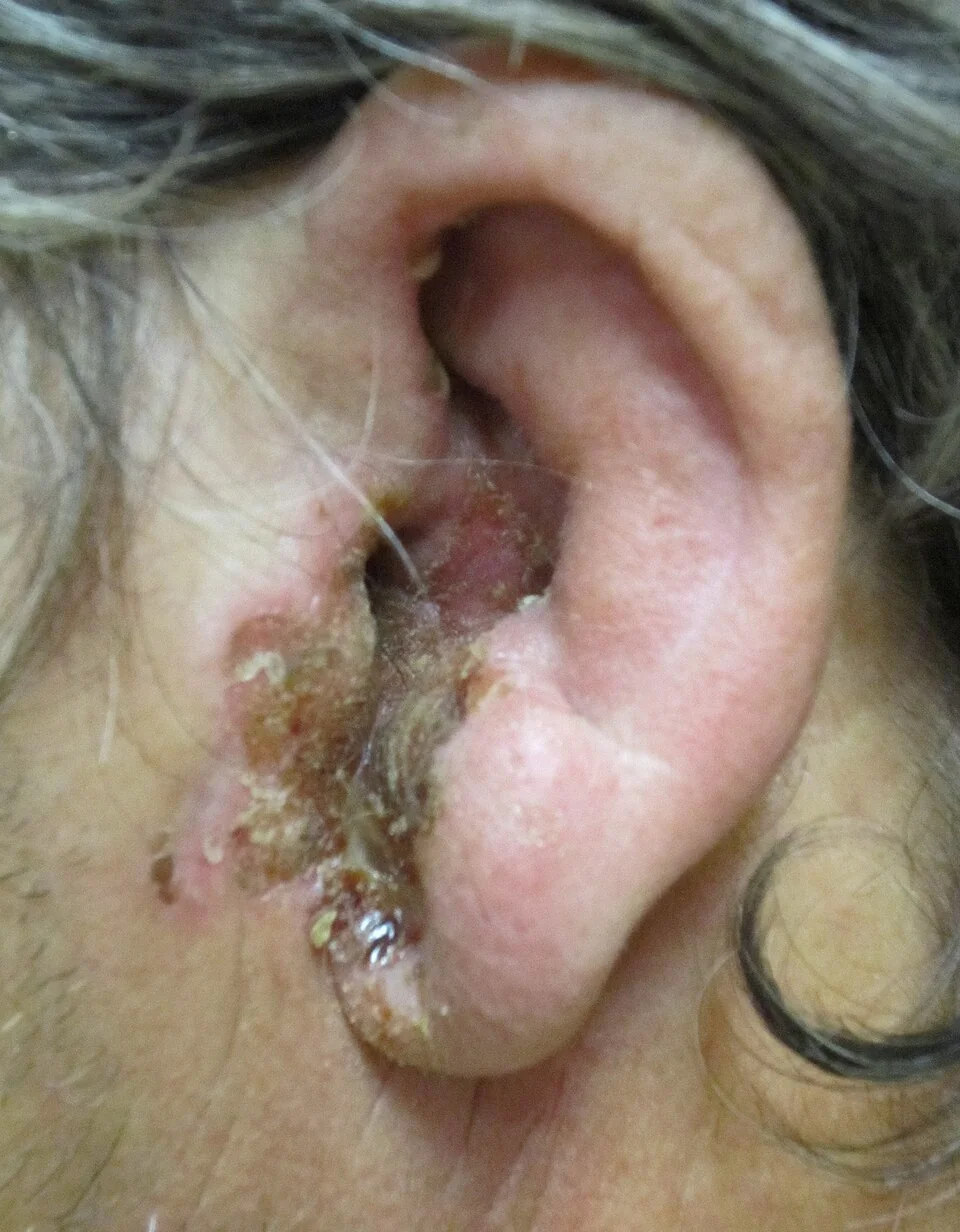
White or black fluffy debris resembling wet tissue paper suggests fungal infection. Granulation tissue on the floor of the canal, particularly at the bony-cartilaginous junction, is a critical finding in a patient with risk factors for malignant otitis externa and should prompt urgent referral rather than routine topical treatment.2
Always examine and document cranial nerve function, particularly the facial nerve, and palpate for tender regional lymphadenopathy. Assess whether the tympanic membrane can be visualised and whether it appears intact, as this affects the choice of topical treatment.
Differential diagnosis
- Acute otitis media (with perforation): discharge but no tragal tenderness; history of preceding URTI
- Malignant otitis externa: severe, persistent pain in a diabetic or immunocompromised patient, granulation tissue, cranial neuropathy
- Furunculosis: a localised, exquisitely tender swelling from an infected hair follicle
- Cholesteatoma: chronic foul-smelling discharge with an attic crust, not an acute painful presentation
- Contact or seborrhoeic dermatitis: itch and scaling without significant pain or discharge
- Foreign body: particularly in children, often unilateral and with a foul odour
Investigations
Otitis externa is a clinical diagnosis and investigations are not needed in typical, uncomplicated cases.1 An ear swab for microscopy, culture and sensitivity is useful when infection is severe, recurrent, resistant to first-line topical treatment, or when fungal infection is suspected.
If malignant otitis externa is suspected, investigate urgently with inflammatory markers (ESR is characteristically very high and useful for monitoring response to treatment), blood glucose/HbA1c, and imaging - CT temporal bones to assess bony erosion, with MRI or technetium/gallium bone scanning to assess the extent of soft tissue and marrow involvement.2
Management
Aural toilet (microsuction or dry mopping of debris and discharge under direct vision) is a key part of treatment, both diagnostically - it allows a clear view of the canal and drum - and therapeutically, since it removes the debris that perpetuates infection and can prevent topical drops from reaching the skin.1
Topical treatment is first line for the great majority of cases. A combined antibiotic and corticosteroid preparation (e.g. neomycin/dexamethasone or gentamicin/hydrocortisone) treats infection and reduces inflammation and itch. Acetic acid 2% spray is a reasonable option for mild cases and works by acidifying the canal. Treatment is typically continued for 7-14 days.1,3
If canal swelling is severe enough to prevent drops reaching the deeper canal, a wick (an ear sponge or ribbon gauze soaked in the topical preparation) is inserted to keep the canal patent and deliver treatment, and is removed or replaced after a few days as the swelling settles.
Oral antibiotics are not routinely required and are reserved for cases with spreading cellulitis beyond the ear, significant systemic illness, or in immunocompromised patients, in which case they should cover Pseudomonas (e.g. ciprofloxacin).3 Otomycosis is treated with an antifungal preparation such as clotrimazole drops after thorough aural toilet, continued for at least 2 weeks.
General advice for all patients includes keeping the ear dry, avoiding swimming until symptoms resolve, and not inserting cotton buds or other objects into the canal, which perpetuates the cycle of trauma and infection.
Malignant (necrotising) otitis externa
Malignant otitis externa is a misleadingly named condition - it is not a malignancy, but an aggressive skull base osteomyelitis that spreads from the external canal into the adjacent temporal bone. It occurs almost exclusively in elderly diabetic patients or others who are immunocompromised, and Pseudomonas aeruginosa is the causative organism in the great majority of cases.2
It should be suspected in anyone with the relevant risk factors who has severe, persistent otalgia (classically worse at night) and otorrhoea that fails to settle with standard topical treatment. Granulation tissue at the bony-cartilaginous junction of the canal floor is a characteristic examination finding. As the infection spreads through the skull base it can cause progressive cranial nerve palsies, most often the facial nerve, followed by the nerves passing through the jugular foramen (IX, X, XI).2
Management requires urgent ENT admission for prolonged intravenous anti-pseudomonal antibiotics (typically a systemic fluoroquinolone or an anti-pseudomonal beta-lactam), tight glycaemic control, and imaging to define the extent of disease and monitor the response to treatment, often continued for 6 weeks or more. Untreated, mortality is significant because of the risk of intracranial extension.
Complications
- Canal stenosis from chronic inflammation and scarring
- Spread of infection to the pinna (perichondritis) or surrounding soft tissue (cellulitis)
- Tympanic membrane perforation with chronic infection
- Malignant otitis externa progressing to skull base osteomyelitis and cranial neuropathy
- Recurrent or chronic otitis externa from repeated water exposure or ongoing trauma
Red flags
Prognosis
Uncomplicated otitis externa resolves within 1-2 weeks with appropriate aural toilet and topical treatment, and most patients need no further follow-up once symptoms settle. Recurrence is common in those who continue to swim regularly or use hearing aids, and such patients benefit from advice on prevention, including drying the ears after swimming and avoiding cotton bud use.1
Malignant otitis externa carries a far more guarded prognosis. Even with prompt treatment, recovery is slow and relapse can occur after treatment is stopped, so patients require close specialist follow-up with clinical and radiological monitoring until resolution is confirmed.2
References
- NICE Clinical Knowledge Summaries (CKS). Otitis externa. 2023. Available here
- Rubin Grandis J, Branstetter BF, Yu VL. The changing face of malignant (necrotising) external otitis: clinical, radiological, and anatomic correlations. Lancet Infectious Diseases. 2004. Available here
- BNF. Ear conditions - infection. Available here
- James Heilman, MD, CC BY 3.0, via Wikimedia Commons. Available here
This article is written for revision and education. It is not clinical guidance and must not be used to make decisions about the care of a patient. Always check current NICE guidance and local protocols.